ARG30270
Loading Control Antibody Panel (Actin, beta Tublin, Histone H3, GAPDH)
Component
| Cat No | Component Name | Host clonality | Reactivity | Application | Package |
|---|---|---|---|---|---|
| ARG65681 | anti-Histone H3 antibody | Mouse mAb | Hu, Ms, Rat | ICC/IF, IHC-P, IP, WB | 50 μg |
| ARG53987 | anti-beta Actin antibody | Mouse mAb | Hu, Ms, Rat, Arabi, C. reinhardtii, Chk, Cucumber, Dm, Fsh, Goat, Hm, Mk, P. pastoris, Pig, Rb, Rice, S. cerevisiae, Yeast, Zfsh | WB | 50 μl |
| ARG54082 | anti-beta Tubulin antibody | Mouse mAb | Hu, Ms, Rat, Goat, Hm, Mk | ICC/IF, WB | 50 μl |
| ARG10112 | anti-GAPDH antibody [6C5] | Mouse mAb | AGMK, Bb, Cat, Chk, Dog, Fsh, Hm, Hu, Mk, Ms, Pig, Rb, Rat, Xenopus laevis, Zfsh | ELISA, ICC/IF, IHC-Fr, WB | 50 μg |
| ARG65350 | Goat anti-Mouse IgG antibody (HRP) | Goat pAb | Ms | ELISA, IHC-P, WB | 50 μl |
Overview
| Product Description | When Western Blot or other experiments are performed, loading controls are required to ensure that (1) the same amount of protein sample is loaded into each lane, (2) protein is transferred from the gel to membrane with equal efficiency across the whole membrane, and (3) antibody incubation and detection is uniform throughout the experimental procedure. Commonly used loading controls include GAPDH, Tubulins, Actin and Histone H3. GAPDH is a key enzyme in the glycolysis. Tubulins and actin are the major building block of microtubules and cytoskeleton. Histone 3 is one of the core proteins in the chromatin. |
|---|---|
| Target Name | Loading Control |
| Alternate Names | Loading Control antibody; GAPDH antibody; beta Actin antibody; beta Tubulin antibody; Histone H3 antibody |
Properties
| Storage Instruction | For continuous use, store undiluted antibody at 2-8°C for up to a week. For long-term storage, aliquot and store at -20°C or below. Storage in frost free freezers is not recommended. Avoid repeated freeze/thaw cycles. Suggest spin the vial prior to opening. The antibody solution should be gently mixed before use. |
|---|---|
| Note | For laboratory research only, not for drug, diagnostic or other use. |
Bioinformation
| Gene Full Name | Antibody Panel for Loading Control (Actin, beta Tublin, Histone H3, GAPDH) |
|---|---|
| Highlight | Related Product: anti-Histone H3 antibody; anti-beta Actin antibody; anti-beta Tubulin antibody; anti-GAPDH antibody; |
Images (21) Click the Picture to Zoom In
-
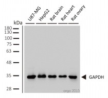
ARG10112 anti-GAPDH antibody [6C5] WB image
Western blot: 1) U87-MG 2) HepG2 3) rat brain 4) rat heart 5) rat ovary stained with ARG10112 anti-GAPDH antibody [6C5] at 1:2000 dilution.
-
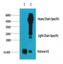
ARG65681 anti-Histone H3 antibody IP image
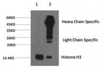
Immunoprecipitation: 1) HeLa cell lysate stained with ARG65681 anti-Histone H3 antibody and 2) IP product immunoprecipitated by ARG65681 anti-Histone H3 antibody at 1:200 dilution.
-
ARG53987 anti-beta Actin antibody WB image
Western blot: 20 µg of HeLa, Mouse brain and Rat brain lysates stained with ARG53987 anti-beta Actin antibody at 1:10000 dilution.
-
ARG54082 anti-beta Tubulin antibody ICC/IF image
Immunofluorescence: 100% Methanol fixed (RT, 10 min) HeLa cells stained with ARG54082 anti-beta Tubulin antibody at 1:500 dilution. Left: primary antibody (green). Middle: DAPI (blue). Right: Merge.
Secondary antibody: ARG55393 Goat anti-Mouse IgG (H+L) antibody (FITC)
-
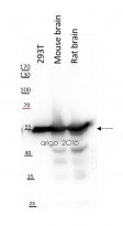
ARG54082 anti-beta Tubulin antibody WB image
Western blot: 20 µg of 293T, Mouse brain and Rat brain lysates stained with ARG54082 anti-beta Tubulin antibody at 1:10000 dilution.
-
ARG65681 anti-Histone H3 antibody ICC/IF image
Immunofluorescence: 100% Methanol fixed (RT, 10 min) HeLa cells stained with ARG65681 anti-Histone H3 antibody at 1:100 dilution. Left: primary antibody (green). Right: Merge (primary antibody and DAPI).
Secondary antibody: ARG55393 Goat anti-Mouse IgG (H+L) antibody (FITC)
-
ARG65681 anti-Histone H3 antibody WB image
Western blot: 20 µg of HeLa, Mouse brain and Rat brain lysates stained with ARG65681 anti-Histone H3 antibody at 1:2000 dilution.
-
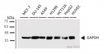
ARG10112 anti-GAPDH antibody [6C5] WB image
Western blot: 1) MCF-7 2) DU-145 3) A549 4) H1299 5) HCT116 6) HepG2 7) HUVEC stained with ARG10112 anti-GAPDH antibody [6C5] at 1:1000 dilution.
-
ARG65681 anti-Histone H3 antibody IHC-P image
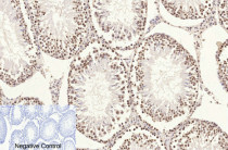
Immunohistochemistry: Paraffin-embedded Rat testis tissue stained with ARG65681 anti-Histone H3 antibody at 1:200 dilution (4°C, overnight). Antigen Retrieval: Boil tissue section in Sodium citrate buffer (pH 6.0) for 20 min. Secondary antibody was diluted at 1:200 (RT, 30 min).
Negative control was used by secondary antibody only.
-
ARG65681 anti-Histone H3 antibody IP image
Immunoprecipitation: 1) HeLa cell lysate stained with ARG65681 anti-Histone H3 antibody and 2) IP product immunoprecipitated by ARG65681 anti-Histone H3 antibody at 1:200 dilution.
-
ARG65681 anti-Histone H3 antibody WB image
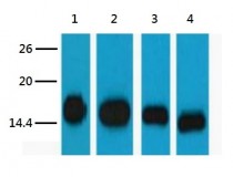
Western blot: 1) HeLa, 2) Raw, 3) Mouse brain tissue, and 4) Rat brain tissue lysates stained with ARG65681 anti-Histone H3 antibody at 1:5000 dilution.
-
ARG10112 anti-GAPDH antibody [6C5] WB image
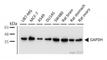
Western blot: 1) U87-MG 2) MCF-7 3) A549 4) DU145 5) SW480 6) rat brain 7) rat stomach 8) rat ovary stained with ARG10112 anti-GAPDH antibody [6C5] at 1:5000 dilution.
-
ARG10112 anti-GAPDH antibody [6C5] WB image
Western blot: pAFs, AcoSV40, and AcoSI-1 stained with ARG10112 anti-GAPDH antibody [6C5] at 1:5000 dilution.
From Michele N Dill et al. PLoS One. (2023), doi: 10.3389/fcell.2022.899869, Fig. 2. C.
-
ARG10112 anti-GAPDH antibody [6C5] WB image
Western blot: Porcine kidney stained with ARG10112 anti-GAPDH antibody [6C5].
From Jianni Huang et al. Front Cell Dev Biol (2022), doi: 10.3389/fcell.2022.899869, Fig. 2. E.
-
ARG10112 anti-GAPDH antibody [6C5] ICC/IF image
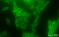
Immunofluorescence: 100% Methanol fixed (RT, 10 min) HeLa cells stained with ARG10112 anti-GAPDH antibody [6C5] (green) at 1:200 dilution.
Secondary antibody: ARG55393 Goat anti-Mouse IgG (H+L) antibody (FITC)
-
ARG10112 anti-GAPDH antibody [6C5] WB image
Western blot: Mouse samples stained with ARG10112 anti-GAPDH antibody [6C5] at 1:1000 dilution.
From Yun-Yun Li et al. Int J Biol Sci (2022), doi: 10.7150/ijbs.68224, Fig. 5. C.
-
ARG10112 anti-GAPDH antibody [6C5] WB image
Western blot: HUVEC stained with ARG10112 anti-GAPDH antibody [6C5].
From Bingzheng Lu et al. Oxid Med Cell Longev (2020), doi: 10.1155/2020/2048210, Fig. 5. B.
-
ARG10112 anti-GAPDH antibody [6C5] IP image
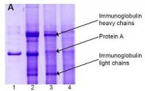
Immunoprecipitation and western blot: 1) GAPDH (1 μg). 2) GAPDH IP from rat heart tissue extract. 3) Only GAPDH preincubated with Protein A Sepharose. 4) Only Protein A Sepharose stained with ARG10112 GAPDH antibody [6C5].
Mixture of protein A-Sepharose with ARG10112 anti-GAPDH and tissue extract was incubated for 30 min at room temperature and precipitated by centrifugation. Pellet was washed with PBS, suspended in reducing electrophoresis sample buffer and heated for 5 minutes at 100ºC. After centrifugation supernatant was loaded on gel and proteins were separated by SDS electrophoresis. -
ARG65350 Goat anti-Mouse IgG antibody (HRP) WB image
Western blot: Rat basolateral amygdala stained with ARG62347 anti-beta Tubulin antibody [BT7R] at 1:1000 dilution, ARG65350 Goat anti-Mouse IgG antibody (HRP) at 1:5000 dilution.
From Guang-Bing Duan et al. CNS Neurosci Ther. (2024), doi: 10.1111/cns.14611, Fig. 4.D.
-
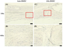
ARG65350 Goat anti-Mouse IgG antibody (HRP) IHC-P image
From Cheng-Feng Chu et al. J Pers Med. (2021), doi: 10.3390/jpm11121326, Fig. 6.
-
ARG10112 anti-GAPDH antibody [6C5] WB image
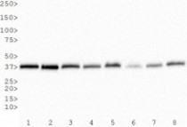
Western Blot: 1) HeLa, 2) NTERA-2, 3) A-431, 4) HepG2, 5) MCF-7, 6) NIH 3T3, 7) PC-12 and 8) COS-7 whole cell lysates stained with anti-GAPDH antibody [6C5] (ARG10112)
Specific References
