APOPTOSIS
APOPTOSIS
|
|
.png)
TOP 3 APOPTOSIS must-haves
Apoptosis Marker
Antibody Duo (Bcl2, Bax)
ARG30268
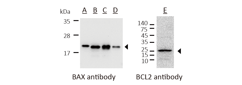
A: MCF7, B: HCT116, C: U87-MG, D: HepG2, E: Jurkat
BAX/BCL2 expression ratio has been identified as an import index to figure out the fate of the cell undergoing apoptosis or anti-apoptosis in variety studies, including cancer study, therapeutic agent searching and therapeutic response evaluation.
Shared reactivity: Human, Mouse Rat
Shared application: WB, IHC-P
Caspase 9 Antibody
ARG54155
.png)
A: HeLa untreated, B: HeLa treated with Staurosporine 1μM (4h)
When a cell receive intra- or extra-cellular death signal, Cytochrome C is released from mitochondria to cytoplasm and trigger Procaspase 9 activation. Detection of the cleaved Caspase 9 represents early indications that a cell is undergoing apoptosis.
Reactivity: Human
Application: WB
Specialty: Recognizing both Caspase 9 precursor and cleaved Caspase 9 protein
PARP (cleaved) Antibody
ARG20041
.png)
A: HeLa untreated, B: HeLa treated with Staurosporine 1μM (4h)
Cleavage of PARP is a pre-requisite for cells who undergo final stage of cell death. The use of anti-cleaved PARP antibody adds a more specific weapon to the arsenal of techniques for confirming apoptosis in tissue specimens.
Reactivity: Human
Application: WB, IHC
Specialty: Recognizing only cleaved PARP p85 protein
