|
Antibodies are often the main ingredient to many widely-used laboratory applications, such as Western Blots (WB), Enzyme-Linked Immunosorbent Assays (ELISA), Immunohistochemistry (IHC), Immunocytochemistry (ICC), Immunoprecipitation (IP), and Flow Cytometry (FACS). In arigo, we mean quality and make it our first priority to provide high quality products to the research community. To this end, we screen our antibodies and validate them with scientific approaches to determine the specificity of our products.
Count on our QC specialists as we bring you through a journey of risk-free antibody shopping experience in arigo.
Western Blotting QC
I Induction of Phosphorylation I
|
|
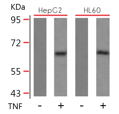
We treat our cell lines or samples with activators for the induction of specific protein phosphorylation.
Extracts from HepG2 and HL60 cells untreated or treated with TNF stained with anti-NFkB p65 (pSer536) antibody ARG51518.
|
- - - - - - - - - - - - - - - - - - - - - - - - - - - - - - - - - - - - - - - - - - - - - - - - - - - - - - - - - - - - - -
I Peptide Blocking I
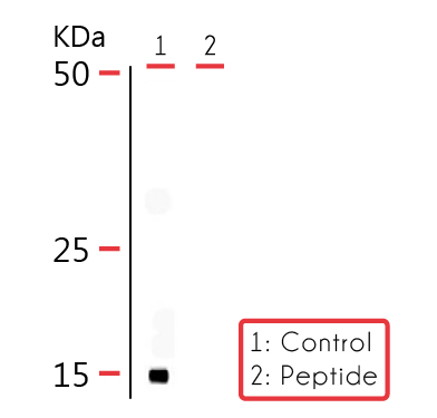 |
To make sure that our antibodies detect a specific epitope of target proteins, we pre-block our samples with peptides before immunolabeling.
Rat mitochondrial lysate stained with ARG52252 COXIV (pSer58) antibody showing specific signal around 17 kDa (Control). Immunolabeling is blocked by the phospho-peptide used as antigen (Peptide).
|
- - - - - - - - - - - - - - - - - - - - - - - - - - - - - - - - - - - - - - - - - - - - - - - - - - - - - - - - - - - - - -
I Phosphatase Treatment I
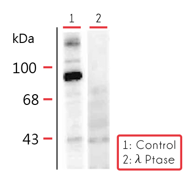 |
We use phosphatase treatments as negative controls for our phospho-specific antibodies.
Forskolin stimulated rat hippocampal lysate showing phospho-specific immunolabeling of the ~95k Dynamin protein phosphorylated at Ser 774 stained with Dynamin (pSer774) antibody (ARG52269).
|
- - - - - - - - - - - - - - - - - - - - - - - - - - - - - - - - - - - - - - - - - - - - - - - - - - - - - - - - - - - - - -
I Inhibitors I
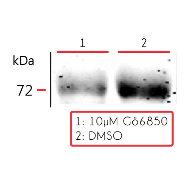 |
Inhibitor treatments help confirming our antibody quality.
Human UACC903 melanoma cell line lysate treated with DMSO as control reagent, showing specific signal at ~ 72 kDa stained with ATF2 (pThr52) antibody (ARG52232).
|
- - - - - - - - - - - - - - - - - - - - - - - - - - - - - - - - - - - - - - - - - - - - - - - - - - - - - - - - - - - - - -
I Multiple Tissues or Cell Lines I
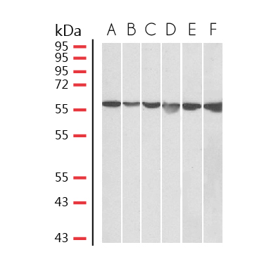 |
Immunolabeling of antibodies in multiple cell lines or tissue samples allow users to make the best choice before purchasing.
HeLa (A), A431 (B), A549 (C), MCF7 (D), Jurkat (E) and K562 (F) lysates (35 µg protein in RIPA buffer) stained with ARG65103 anti-GPI / Neuroleukin (aa81-93) antibody at 0.05 µg/ml dilution.
|
- - - - - - - - - - - - - - - - - - - - - - - - - - - - - - - - - - - - - - - - - - - - - - - - - - - - - - - - - - - - - -
I Overexpression I
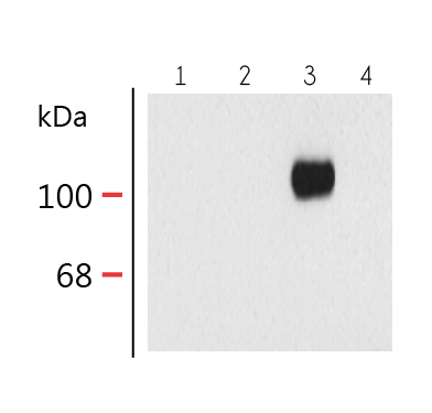 |
We validate our antibodies specific to certain variant or isoform by comparing WB data in various overexpression lines.
10 μg of HEK 293 cells expressing: Lane 1 - HEK cells without NR1 expression (Mock); Lane 2 - NR1 subunit containing only the C2 Insert; Lane 3 - NR1 subunit containing the C1 and C2' Insert; Lane 4 - NR1 subunit containing the N1 and C2' Insert stained with ARG52353 anti-NDMAR1, Splice Variant C1 antibody showing specific immunolabeling of the ~120k NR1 subunit of the NMDA receptor containing the C1 splice variant insert.
|
Click here for more primary antibodies
Immunocytochemistry QC
I Cellular Localization I
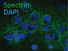 |
We make sure that there is precise cellular localization of immunolabeling by counter-staining with certain organelle markers.
Cultured rat neurons and glia stained with ARG52218 Alpha II Spectrin antibody [3D7] showing axonal and dendritic staining of alpha II spectrin (green) revealing the submembraneous cytoskeleton and DNA (blue).
|
- - - - - - - - - - - - - - - - - - - - - - - - - - - - - - - - - - - - - - - - - - - - - - - - - - - - - - - - - - - - - -
I Cell Type Confirmation I
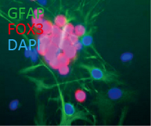 |
We scientifically verify the specificity of our antibodies by counter-staining with markers of different cell types.
Cultured rat neurons stained with ARG52283 anti-FOX3 (NeuN) antibody [1B7] showing strong nuclear and distal cytoplasmic staining of FOX3 in red and the complete absence of astrocyte staining, which are stained green stained with ARG52313 anti-GFAP antibody.
|
- - - - - - - - - - - - - - - - - - - - - - - - - - - - - - - - - - - - - - - - - - - - - - - - - - - - - - - - - - - - - -
I Induction of Phosphorylation I
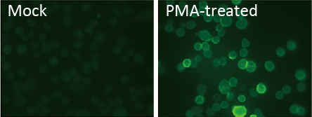 |
We treat our cell lines or samples with activators for the induction of specific protein phosphorylation.
Medial nucleus of the trapezoid body (MNTB) cells stained with ARG52399 Kv3.1 (p503) antibody. The left panel shows control cells. The right panel shows cells that have been exposed to the protein kinase C activator PMA.
|
Click here for more primary antibodies
Immunohistochemistry QC
I Peptide Blocking I
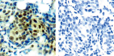 |
To make sure that our antibodies detect a specific epitope of target proteins, we pre-block our samples with peptides before immunolabeling.
Paraffin-embedded human breast carcinoma tissue stained with anti-p44/42 MAP Kinase (pThr202) antibody ARG51732 (left) or the same antibody preincubated with blocking peptide (right).
|
- - - - - - - - - - - - - - - - - - - - - - - - - - - - - - - - - - - - - - - - - - - - - - - - - - - - - - - - - - - - - -
I Expression Patterns I
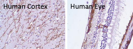 |
The expression patterns of target protein in certain tissue types are carefully examined and validated.
Human Cerebel Cortex (left) and human eye (right) stained with Glial Fibrillary Acidic Protein (GFAP) antibody [SP78] (ARG53074). Note the different expression patterns in these two sample types.
|
Click here for more primary antibodies
|