ARG65681
anti-Histone H3 antibody
anti-Histone H3 antibody for ICC/IF,IHC-Formalin-fixed paraffin-embedded sections,Immunoprecipitation,Western blot and Human,Mouse,Rat
Controls and Markers antibody; Gene Regulation antibody; Loading Control antibody; Loading Control antibody for Nuclear Fractions; Organelle Marker antibody for Nucleus; Nuclear translocation Study antibody; CARM1 mediated histone arginine methylation antibody; Cell Cycle Study antibody; Polycomb Complexes antibody

Overview
| Product Description | Mouse Monoclonal antibody recognizes Histone H3 |
|---|---|
| Tested Reactivity | Hu, Ms, Rat |
| Tested Application | ICC/IF, IHC-P, IP, WB |
| Host | Mouse |
| Clonality | Monoclonal |
| Isotype | IgG2b, kappa |
| Target Name | Histone H3 |
| Antigen Species | Human |
| Immunogen | Recombinant Protein of Human Histone H3 |
| Conjugation | Un-conjugated |
| Alternate Names | Histone H3/f; Histone H3.1; Histone H3/d; Histone H3/b; Histone H3/c; Histone H3/a; Histone H3/l; Histone H3/j; Histone H3/k; Histone H3/h; H3/A; H3FA; Histone H3/i |
Application Instructions
| Application Suggestion |
|
||||||||||
|---|---|---|---|---|---|---|---|---|---|---|---|
| Application Note | IHC-P: Antigen Retrieval: Boil tissue section in Sodium citrate buffer (pH 6.0) for 20 min. * The dilutions indicate recommended starting dilutions and the optimal dilutions or concentrations should be determined by the scientist. |
Properties
| Form | Liquid |
|---|---|
| Purification | Affinity purification with immunogen. |
| Buffer | PBS (pH 7.4), 0.02% Sodium azide and 50% Glycerol |
| Preservative | 0.02% Sodium azide |
| Stabilizer | 50% Glycerol |
| Concentration | 1 mg/ml |
| Storage Instruction | For continuous use, store undiluted antibody at 2-8°C for up to a week. For long-term storage, aliquot and store at -20°C. Storage in frost free freezers is not recommended. Avoid repeated freeze/thaw cycles. Suggest spin the vial prior to opening. The antibody solution should be gently mixed before use. |
| Note | For laboratory research only, not for drug, diagnostic or other use. |
Bioinformation
| Database Links | |
|---|---|
| Gene Symbol | HIST1H3A |
| Gene Full Name | histone cluster 1, H3a |
| Background | Histones are basic nuclear proteins that are responsible for the nucleosome structure of the chromosomal fiber in eukaryotes. This structure consists of approximately 146 bp of DNA wrapped around a nucleosome, an octamer composed of pairs of each of the four core histones (H2A, H2B, H3, and H4). The chromatin fiber is further compacted through the interaction of a linker histone, H1, with the DNA between the nucleosomes to form higher order chromatin structures. This gene is intronless and encodes a replication-dependent histone that is a member of the histone H3 family. Transcripts from this gene lack polyA tails; instead, they contain a palindromic termination element. This gene is found in the large histone gene cluster on chromosome 6p22-p21.3. [provided by RefSeq, Aug 2015] |
| Function | Core component of nucleosome. Nucleosomes wrap and compact DNA into chromatin, limiting DNA accessibility to the cellular machineries which require DNA as a template. Histones thereby play a central role in transcription regulation, DNA repair, DNA replication and chromosomal stability. DNA accessibility is regulated via a complex set of post-translational modifications of histones, also called histone code, and nucleosome remodeling. [UniProt] |
| Highlight | Related Antibody Duos and Panels: ARG30259 Loading Controls for Cytoplasmic / Nuclear Fractions Antibody Panel ARG30267 Organelle Marker, Cytoplasm, Nucleus Antibody Duo (Histone, GAPDH) ARG30270 Loading Control Antibody Panel (Actin, beta Tublin, Histone H3, GAPDH) Related products: Histone H3 antibodies; Histone H3 Duos / Panels; Related news: Is it a potential cancer therapy by preventing acetate capture? |
| Research Area | Controls and Markers antibody; Gene Regulation antibody; Loading Control antibody; Loading Control antibody for Nuclear Fractions; Organelle Marker antibody for Nucleus; Nuclear translocation Study antibody; CARM1 mediated histone arginine methylation antibody; Cell Cycle Study antibody; Polycomb Complexes antibody |
| Calculated MW | 15 kDa |
| PTM | Acetylation is generally linked to gene activation. Acetylation on Lys-10 (H3K9ac) impairs methylation at Arg-9 (H3R8me2s). Acetylation on Lys-19 (H3K18ac) and Lys-24 (H3K24ac) favors methylation at Arg-18 (H3R17me). Acetylation at Lys-123 (H3K122ac) by EP300/p300 plays a central role in chromatin structure: localizes at the surface of the histone octamer and stimulates transcription, possibly by promoting nucleosome instability. Citrullination at Arg-9 (H3R8ci) and/or Arg-18 (H3R17ci) by PADI4 impairs methylation and represses transcription. Asymmetric dimethylation at Arg-18 (H3R17me2a) by CARM1 is linked to gene activation. Symmetric dimethylation at Arg-9 (H3R8me2s) by PRMT5 is linked to gene repression. Asymmetric dimethylation at Arg-3 (H3R2me2a) by PRMT6 is linked to gene repression and is mutually exclusive with H3 Lys-5 methylation (H3K4me2 and H3K4me3). H3R2me2a is present at the 3' of genes regardless of their transcription state and is enriched on inactive promoters, while it is absent on active promoters. Methylation at Lys-5 (H3K4me), Lys-37 (H3K36me) and Lys-80 (H3K79me) are linked to gene activation. Methylation at Lys-5 (H3K4me) facilitates subsequent acetylation of H3 and H4. Methylation at Lys-80 (H3K79me) is associated with DNA double-strand break (DSB) responses and is a specific target for TP53BP1. Methylation at Lys-10 (H3K9me) and Lys-28 (H3K27me) are linked to gene repression. Methylation at Lys-10 (H3K9me) is a specific target for HP1 proteins (CBX1, CBX3 and CBX5) and prevents subsequent phosphorylation at Ser-11 (H3S10ph) and acetylation of H3 and H4. Methylation at Lys-5 (H3K4me) and Lys-80 (H3K79me) require preliminary monoubiquitination of H2B at 'Lys-120'. Methylation at Lys-10 (H3K9me) and Lys-28 (H3K27me) are enriched in inactive X chromosome chromatin. Monomethylation at Lys-57 (H3K56me1) by EHMT2/G9A in G1 phase promotes interaction with PCNA and is required for DNA replication. Phosphorylated at Thr-4 (H3T3ph) by GSG2/haspin during prophase and dephosphorylated during anaphase. Phosphorylation at Ser-11 (H3S10ph) by AURKB is crucial for chromosome condensation and cell-cycle progression during mitosis and meiosis. In addition phosphorylation at Ser-11 (H3S10ph) by RPS6KA4 and RPS6KA5 is important during interphase because it enables the transcription of genes following external stimulation, like mitogens, stress, growth factors or UV irradiation and result in the activation of genes, such as c-fos and c-jun. Phosphorylation at Ser-11 (H3S10ph), which is linked to gene activation, prevents methylation at Lys-10 (H3K9me) but facilitates acetylation of H3 and H4. Phosphorylation at Ser-11 (H3S10ph) by AURKB mediates the dissociation of HP1 proteins (CBX1, CBX3 and CBX5) from heterochromatin. Phosphorylation at Ser-11 (H3S10ph) is also an essential regulatory mechanism for neoplastic cell transformation. Phosphorylated at Ser-29 (H3S28ph) by MAP3K20 isoform 1, RPS6KA5 or AURKB during mitosis or upon ultraviolet B irradiation. Phosphorylation at Thr-7 (H3T6ph) by PRKCB is a specific tag for epigenetic transcriptional activation that prevents demethylation of Lys-5 (H3K4me) by LSD1/KDM1A. At centromeres, specifically phosphorylated at Thr-12 (H3T11ph) from prophase to early anaphase, by DAPK3 and PKN1. Phosphorylation at Thr-12 (H3T11ph) by PKN1 is a specific tag for epigenetic transcriptional activation that promotes demethylation of Lys-10 (H3K9me) by KDM4C/JMJD2C. Phosphorylation at Thr-12 (H3T11ph) by chromatin-associated CHEK1 regulates the transcription of cell cycle regulatory genes by modulating acetylation of Lys-10 (H3K9ac). Phosphorylation at Tyr-42 (H3Y41ph) by JAK2 promotes exclusion of CBX5 (HP1 alpha) from chromatin. Monoubiquitinated by RAG1 in lymphoid cells, monoubiquitination is required for V(D)J recombination (By similarity). Ubiquitinated by the CUL4-DDB-RBX1 complex in response to ultraviolet irradiation. This may weaken the interaction between histones and DNA and facilitate DNA accessibility to repair proteins. Lysine deamination at Lys-5 (H3K4all) to form allysine is mediated by LOXL2. Allysine formation by LOXL2 only takes place on H3K4me3 and results in gene repression (PubMed:22483618). Crotonylation (Kcr) is specifically present in male germ cells and marks testis-specific genes in post-meiotic cells, including X-linked genes that escape sex chromosome inactivation in haploid cells. Crotonylation marks active promoters and enhancers and confers resistance to transcriptional repressors. It is also associated with post-meiotically activated genes on autosomes. |
Images (4) Click the Picture to Zoom In
-
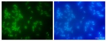
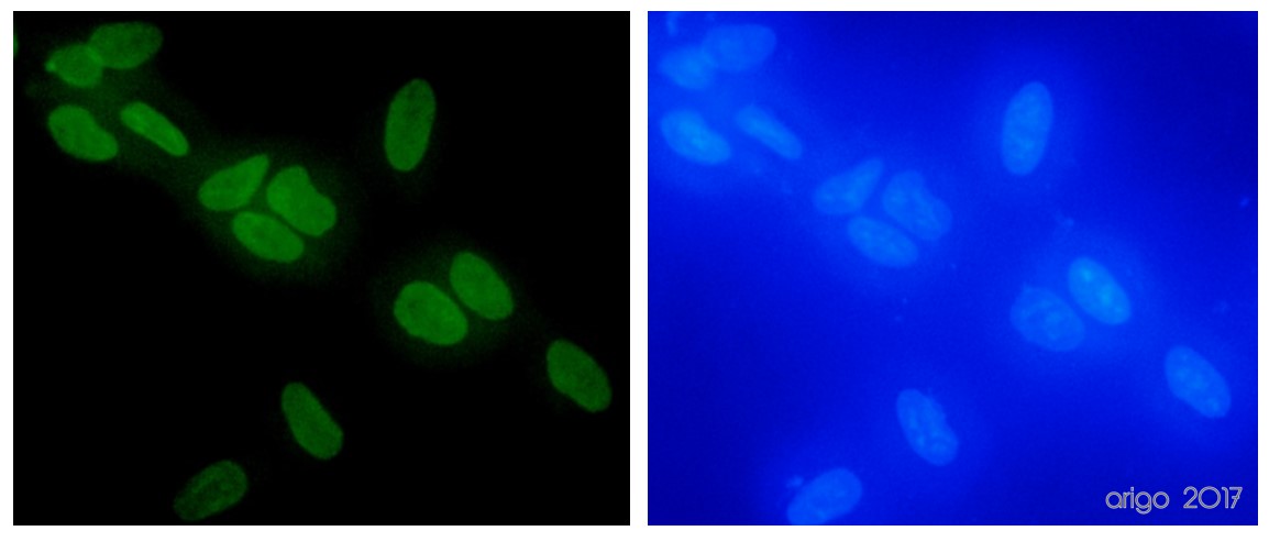
ARG65681 anti-Histone H3 antibody ICC/IF image
Immunofluorescence: 100% Methanol fixed (RT, 10 min) HeLa cells stained with ARG65681 anti-Histone H3 antibody at 1:100 dilution. Left: primary antibody (green). Right: Merge (primary antibody and DAPI).
Secondary antibody: ARG55393 Goat anti-Mouse IgG (H+L) antibody (FITC)
-
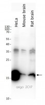
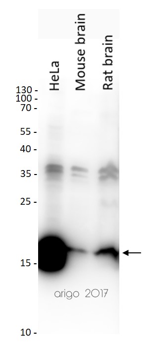
ARG65681 anti-Histone H3 antibody WB image
Western blot: 20 µg of HeLa, Mouse brain and Rat brain lysates stained with ARG65681 anti-Histone H3 antibody at 1:2000 dilution.
-
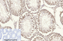
ARG65681 anti-Histone H3 antibody IHC-P image
Immunohistochemistry: Paraffin-embedded Rat testis tissue stained with ARG65681 anti-Histone H3 antibody at 1:200 dilution (4°C, overnight). Antigen Retrieval: Boil tissue section in Sodium citrate buffer (pH 6.0) for 20 min. Secondary antibody was diluted at 1:200 (RT, 30 min).
Negative control was used by secondary antibody only.
-
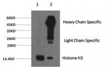
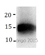
ARG65681 anti-Histone H3 antibody IP image
Immunoprecipitation: 1) HeLa cell lysate stained with ARG65681 anti-Histone H3 antibody and 2) IP product immunoprecipitated by ARG65681 anti-Histone H3 antibody at 1:200 dilution.


Customer's Feedback
Excellent
Review for anti-Histone H3 antibody
Application:WB
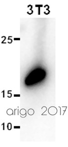
Sample:3T3
Sample Loading Amount:20 µg
Primary Antibody Dilution Factor:1:2000
Primary Antibody Incubation Time:overnight
Primary Antibody Incubation Temperature:4 ºC

Excellent
Review for anti-Histone H3 antibody
Application:IF/ICC
Sample:HeLa
Fixation Buffer:100% Methanol
Fixation Time:10 min
Fixation Temperature:RT ºC
Permeabilization Buffer:0.1% Triton X-100
Primary Antibody Dilution Factor:1:100
Primary Antibody Incubation Time:overnight
Primary Antibody Incubation Temperature:4 ºC
Conjugation of Secondary Antibody:FITC
Excellent
Review for anti-Histone H3 antibody
Application:WB
Sample:HeLa, Mouse brain and Rat brain
Sample Loading Amount:20 µg
Primary Antibody Dilution Factor:1:2000
Primary Antibody Incubation Time:overnight
Primary Antibody Incubation Temperature:4 ºC
Excellent
Review for anti-Histone H3 antibody
Application:WB
Sample:293T
Sample Loading Amount:20 µg
Primary Antibody Dilution Factor:1:500
Primary Antibody Incubation Time:overnight
Primary Antibody Incubation Temperature:4 ºC
Specific References
Gut microbiome dysbiosis contributes to abdominal aortic aneurysm by promoting neutrophil extracellular trap formation
IHC-P / Mouse


