ARG65863
anti-HMGB1 antibody [SQab1711]
anti-HMGB1 antibody [SQab1711] for ELISA,Flow cytometry,ICC/IF,IHC-Formalin-fixed paraffin-embedded sections,Western blot and Human,Mouse,Rat

Overview
| Product Description | Mouse Monoclonal antibody [SQab1711] recognizes HMGB1 |
|---|---|
| Tested Reactivity | Hu, Ms, Rat |
| Tested Application | ELISA, FACS, ICC/IF, IHC-P, WB |
| Host | Mouse |
| Clonality | Monoclonal |
| Clone | SQab1711 |
| Isotype | IgG2b |
| Target Name | HMGB1 |
| Antigen Species | Human |
| Immunogen | OVA-conjugated synthetic peptide of HMGB1. |
| Conjugation | Un-conjugated |
| Alternate Names | HMG-1; High mobility group protein B1; High mobility group protein 1; HMG1; SBP-1; HMG3 |
Application Instructions
| Application Suggestion |
|
||||||||||||
|---|---|---|---|---|---|---|---|---|---|---|---|---|---|
| Application Note | IHC-P: Antigen Retrieval: Boil tissue section in 1X EDTA buffer pH9.0 for 10 -20 min followed by cooling at RT for 20 min. * The dilutions indicate recommended starting dilutions and the optimal dilutions or concentrations should be determined by the scientist. |
Properties
| Form | Liquid |
|---|---|
| Purification | Purification with Protein G. |
| Buffer | PBS (pH 7.4) and 0.01% Thimerosal. |
| Preservative | 0.01% Thimerosal. |
| Concentration | 1 mg/ml |
| Storage Instruction | For continuous use, store undiluted antibody at 2-8°C for up to a week. For long-term storage, aliquot and store at -20°C or below. Storage in frost free freezers is not recommended. Avoid repeated freeze/thaw cycles. Suggest spin the vial prior to opening. The antibody solution should be gently mixed before use. |
| Note | For laboratory research only, not for drug, diagnostic or other use. |
Bioinformation
| Database Links | |
|---|---|
| Gene Symbol | HMGB1 |
| Gene Full Name | high mobility group box 1 |
| Background | HMGB1 is a protein that belongs to the High Mobility Group-box superfamily. The encoded non-histone, nuclear DNA-binding protein regulates transcription, and is involved in organization of DNA. This protein plays a role in several cellular processes, including inflammation, cell differentiation and tumor cell migration. Multiple pseudogenes of this gene have been identified. Alternative splicing results in multiple transcript variants that encode the same protein. [provided by RefSeq, Sep 2015] |
| Function | HMGB1 is a DNA binding protein. It associates with chromatin and has the ability to bend DNA. Binds preferentially single-stranded DNA. Involved in V(D)J recombination by acting as a cofactor of the RAG complex. Acts by stimulating cleavage and RAG protein binding at the 23 bp spacer of conserved recombination signal sequences (RSS). [UniProt] |
| Highlight | Related Antibody Duos and Panels: ARG30343 GSDME-mediated Pyroptosis Antibody Panel Related products: HMGB1 antibodies; HMGB1 ELISA Kits; HMGB1 Duos / Panels; HMGB1 recombinant proteins; Anti-Mouse IgG secondary antibodies; Related news: HMGB1, a biomarker and therapeutic target in COVID-19 Total solution for HMGB1 research HMGB1 in inflammation Inflammatory Cytokines HMGB1 ELISA Kit for your research Detecting the DAMPs in cancer therapy by HMGB1 ELISA kit New HMGB1 neutralizing antibody is released Detecting exosomal HMGB1 for ICD research Related poster download: HMGB2 Pathway.pdf |
| Calculated MW | 25 kDa |
| PTM | Phosphorylated at serine residues. Phosphorylation in both NLS regions is required for cytoplasmic translocation followed by secretion (PubMed:17114460). Acetylated on multiple sites upon stimulation with LPS (PubMed:22801494). Acetylation on lysine residues in the nuclear localization signals (NLS 1 and NLS 2) leads to cytoplasmic localization and subsequent secretion (By similarity). Acetylation on Lys-3 results in preferential binding to DNA ends and impairs DNA bending activity (By similarity). Reduction/oxidation of cysteine residues Cys-23, Cys-45 and Cys-106 and a possible intramolecular disulfide bond involving Cys-23 and Cys-45 give rise to different redox forms with specific functional activities in various cellular compartments: 1- fully reduced HMGB1 (HMGB1C23hC45hC106h), 2- disulfide HMGB1 (HMGB1C23-C45C106h) and 3- sulfonyl HMGB1 (HMGB1C23soC45soC106so). Poly-ADP-ribosylated by PARP1 when secreted following stimulation with LPS (By similarity). In vitro cleavage by CASP1 is liberating a HMG box 1-containing peptide which may mediate immunogenic activity; the peptide antagonizes apoptosis-induced immune tolerance (PubMed:24474694). Can be proteolytically cleaved by a thrombin:thrombomodulin complex. |
Images (11) Click the Picture to Zoom In
-
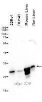
ARG65863 anti-HMGB1 antibody [SQab1711] WB image
Western blot: 20 µg of 22Rv1, DU145, Mouse liver and Rat liver lysates stained with ARG65863 anti-HMGB1 antibody [SQab1711] at 1:5000 dilution.
-
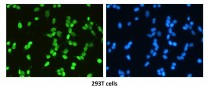
ARG65863 anti-HMGB1 antibody [SQab1711] ICC/IF image
Immunofluorescence: 293T cells stained with ARG65863 anti-HMGB1 antibody [SQab1711] at 5 μg/ml dilution.
-
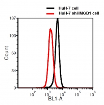
ARG65863 anti-HMGB1 antibody [SQab1711] FACS image
Flow Cytometry: HuH-7 cell and HuH-7 shHMGB1 cell stained with ARG65863 anti-HMGB1 antibody [SQab1711] at 1 µg/ml dilution.
-
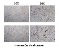
ARG65863 anti-HMGB1 antibody [SQab1711] IHC-P image
Immunohistochemistry: Paraffin-embedded Human cervical cancer stained with ARG65863 anti-HMGB1 antibody [SQab1711] at 10 μg/ml dilution.
-
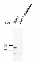
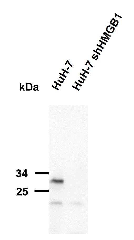
ARG65863 anti-HMGB1 antibody [SQab1711] WB image
Western blot: 50 µg of 1) HuH-7 and 2) HuH-7 shHMGB1 cell lysates stained with ARG65863 anti-HMGB1 antibody [SQab1711] at 1:5000 dilution.
-
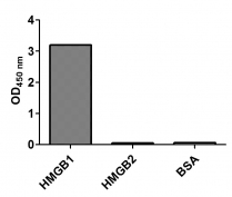
ARG65863 anti-HMGB1 antibody [SQab1711] ELISA image
ELISA: ARG65863 anti-HMGB1 antibody [SQab1711] at 1:5000 were used for detecting HMGB1. 5 µg/ml of HMGB1, HMGB2 and BSA proteins were coated onto ELISA plate.
-
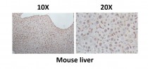
ARG65863 anti-HMGB1 antibody [SQab1711] IHC-P image
Immunohistochemistry: Paraffin-embedded Mouse liver stained with ARG65863 anti-HMGB1 antibody [SQab1711] at 10 μg/ml dilution.
-
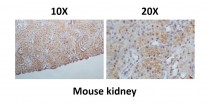
ARG65863 anti-HMGB1 antibody [SQab1711] IHC-P image
Immunohistochemistry: Paraffin-embedded Mouse kidney stained with ARG65863 anti-HMGB1 antibody [SQab1711] at 10 μg/ml dilution.
-
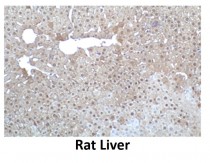
ARG65863 anti-HMGB1 antibody [SQab1711] IHC-P image
Immunohistochemistry: Paraffin-embedded Rat liver stained with ARG65863 anti-HMGB1 antibody [SQab1711] at 10 μg/ml dilution.
-
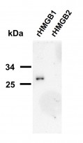
ARG65863 anti-HMGB1 antibody [SQab1711] WB image
Western blot: 1) 50 ng of HMGB1 (Purified from E. coli) and 2) 100 ng of HMGB2 (Purified from E.coli) stained with ARG65863 anti-HMGB1 antibody [SQab1711] at 1:5000 dilution.
-
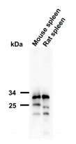
ARG65863 anti-HMGB1 antibody [SQab1711] WB image
Western blot: 20 µg of 1) Mouse spleen and 2) Rat spleen lysates stained with ARG65863 anti-HMGB1 antibody [SQab1711] at 1:5000 dilution.


Customer's Feedback
Excellent

Review for anti-HMGB1 antibody [SQab1711]
Application:WB
Sample:MCF7
Sample Loading Amount:20 µg
Primary Antibody Dilution Factor:1:5000
Primary Antibody Incubation Time:overnight
Primary Antibody Incubation Temperature:4 ºC

Specific References
Tumor cell-released autophagosomes (TRAPs) induce PD-L1-decorated NETs that suppress T-cell function to promote breast cancer pulmonary metastasis
FACS / Mouse


Expression and clinical significance of high mobility group protein B1 in patients with hemophagocytic lymphohistiocytosis.
ELISA / Human


