ARG67000
anti-CD3 antibody
anti-CD3 antibody for Flow cytometry,ICC/IF,IHC-Formalin-fixed paraffin-embedded sections,Western blot and Human,Mouse
Cancer antibody; Developmental Biology antibody; Immune System antibody; Lymphocyte Marker antibody; Inflammatory Cell Marker antibody; T-cell Marker antibody; T-cell infiltration Study antibody; Tumor-infiltrating Lymphocyte Study antibody
Overview
| Product Description | Rabbit Polyclonal antibody recognizes CD3 |
|---|---|
| Tested Reactivity | Hu, Ms |
| Predict Reactivity | Rat |
| Tested Application | FACS, ICC/IF, IHC-P, WB |
| Host | Rabbit |
| Clonality | Polyclonal |
| Isotype | IgG |
| Target Name | CD3 |
| Antigen Species | Human |
| Immunogen | Synthetic peptide corresponding to the intracellular domain of Human CD3 epsilon. |
| Conjugation | Un-conjugated |
| Alternate Names | T-cell surface antigen T3/Leu-4 epsilon chain; T3E; TCRE; T-cell surface glycoprotein CD3 epsilon chain; IMD18; CD antigen CD3e |
Application Instructions
| Application Suggestion |
|
||||||||||
|---|---|---|---|---|---|---|---|---|---|---|---|
| Application Note | IHC-P: Antigen Retrieval: Boil tissue section in Citrate buffer (pH 6.0). * The dilutions indicate recommended starting dilutions and the optimal dilutions or concentrations should be determined by the scientist. |
||||||||||
| Positive Control | Jurkat; Thymus tissue; Spleen; Tonsil carcinoma | ||||||||||
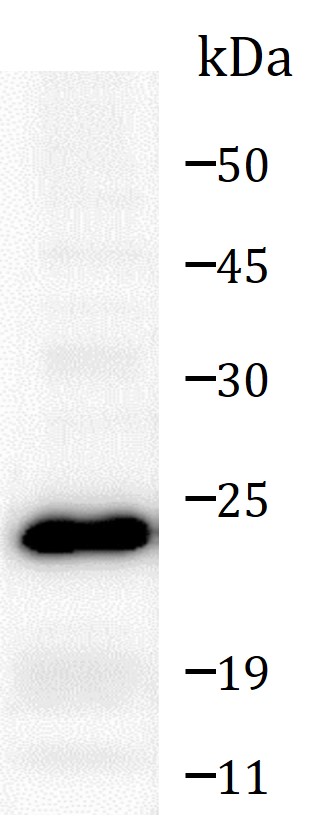
| Observed Size | 23 kDa |
Properties
| Form | Liquid |
|---|---|
| Purification | Affinity purified. |
| Buffer | 100 mM Tris Glycine (pH 7.0), 0.025% ProClin 300 and 20% Glycerol. |
| Preservative | 0.025% ProClin 300 |
| Stabilizer | 20% Glycerol |
| Storage Instruction | For continuous use, store undiluted antibody at 2-8°C for up to a week. For long-term storage, aliquot and store at -20°C. Storage in frost free freezers is not recommended. Avoid repeated freeze/thaw cycles. Suggest spin the vial prior to opening. The antibody solution should be gently mixed before use. |
| Note | For laboratory research only, not for drug, diagnostic or other use. |
Bioinformation
| Database Links |
Swiss-port # P07766 Human T-cell surface glycoprotein CD3 epsilon chain Swiss-port # P22646 Mouse T-cell surface glycoprotein CD3 epsilon chain |
|---|---|
| Gene Symbol | CD3E |
| Gene Full Name | CD3e molecule, epsilon (CD3-TCR complex) |
| Background | CD3 subunit complex is crucial in transducing antigen-recognition signals into the cytoplasm of T cells and in regulating the cell surface expression of the TCR complex. T cell activation through the antigen receptor (TCR) involves the cytoplasmic tails of the CD3 subunits CD3 gamma, CD3 delta, CD3 epsilon and CD3 zeta. These CD3 subunits are structurally related members of the immunoglobulins superfamily encoded by closely linked genes on human chromosome 11. The CD3 components have long cytoplasmic tails that associate with cytoplasmic signal transduction molecules. This association is mediated at least in part by a double tyrosine-based motif present in a single copy in the CD3 subunits. CD3 may play a role in TCR-induced growth arrest, cell survival and proliferation. |
| Function | CD3: Part of the TCR-CD3 complex present on T-lymphocyte cell surface that plays an essential role in adaptive immune response. When antigen presenting cells (APCs) activate T-cell receptor (TCR), TCR-mediated signals are transmitted across the cell membrane by the CD3 chains CD3D, CD3E, CD3G and CD3Z. All CD3 chains contain immunoreceptor tyrosine-based activation motifs (ITAMs) in their cytoplasmic domain. Upon TCR engagement, these motifs become phosphorylated by Src family protein tyrosine kinases LCK and FYN, resulting in the activation of downstream signaling pathways (PubMed:2470098). In addition of this role of signal transduction in T-cell activation, CD3E plays an essential role in correct T-cell development. Initiates the TCR-CD3 complex assembly by forming the two heterodimers CD3D/CD3E and CD3G/CD3E. Participates also in internalization and cell surface down-regulation of TCR-CD3 complexes via endocytosis sequences present in CD3E cytosolic region (PubMed:10384095, PubMed:26507128). [UniProt] |
| Highlight | Related Antibody Duos and Panels: ARG30302 T-cell infiltration Antibody Duo ARG30325 Inflammatory Cell Antibody Panel ARG30334 Tumor-infiltrating Lymphocyte Antibody Panel Related products: CD3 antibodies; CD3 ELISA Kits; CD3 Duos / Panels; CD3 recombinant proteins; Anti-Rabbit IgG secondary antibodies; Related news: More effective cocktail therapy for cancer immune evasion Cholesterol, the weakness of anaplastic large cell lymphoma (ALCL) New antibody panels and duos for Tumor immune microenvironment Tumor-Infiltrating Lymphocytes (TILs) Exploring Antiviral Immune Response |
| Research Area | Cancer antibody; Developmental Biology antibody; Immune System antibody; Lymphocyte Marker antibody; Inflammatory Cell Marker antibody; T-cell Marker antibody; T-cell infiltration Study antibody; Tumor-infiltrating Lymphocyte Study antibody |
| Calculated MW | 23 kDa |
Images (5) Click the Picture to Zoom In
-
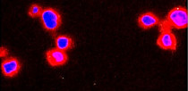
ARG67000 anti-CD3 antibody ICC/IF image
Immunofluorescence: Jurkat cells were fixed with 4% paraformaldehyde at RT for 10 min, permeabilized with 0.1% NP-40 at RT for 10 min and then cells were blocked with 5% BSA at RT for 30 min and stained with ARG67000 anti-CD3 antibody (red) at 1:200 dilution at 4°C for overnight. DAPI (blue) was used for nuclear staining.
-
_image_210_205.jpg)
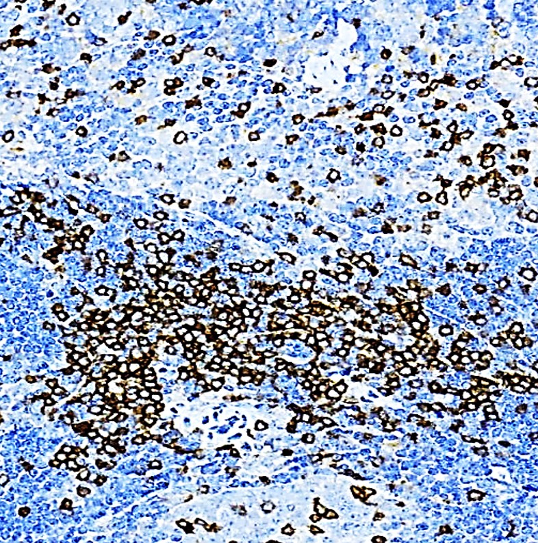
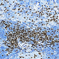
ARG67000 anti-CD3 antibody IHC-P(Human) image
Immunohistochemistry: Paraffin-embedded Human Tonsil carcinoma tissue stained with ARG67000 anti-CD3 antibody at 1:200 dilution. Antigen Retrieval: Boil tissue section in Citrate buffer (pH 6.0).
-
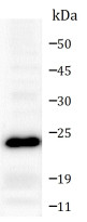
ARG67000 anti-CD3 antibody WB image
Western blot: 40 µg of Jurkat cell and Mouse Thymus tissue lysate stained with ARG67000 anti-CD3 antibody at 1:1000 dilution, overnight at 4°C.
-
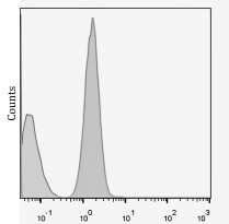
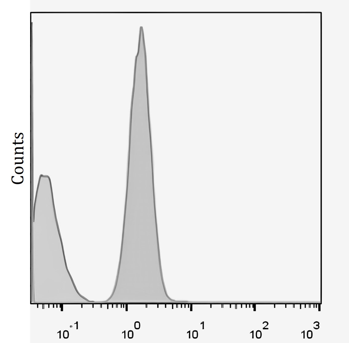
ARG67000 anti-CD3 antibody FACS image
Flow Cytometry: Human whole blood stained with ARG67000 anti-CD3 antibody at 1:100 dilution (grey area).
-
ARG67000 anti-CD3 antibody IHC-P(Mouse) image
Immunohistochemistry: Paraffin-embedded Mouse spleen tissue stained with ARG67000 anti-CD3 antibody at 1:200 dilution. Antigen Retrieval: Boil tissue section in Citrate buffer (pH 6.0).
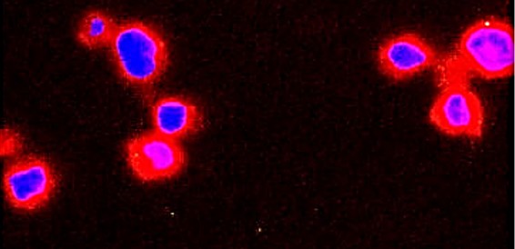
_image.jpg)