ARG30131
Postsynaptic Receptor Antibody Panel (NMDAR2A, NMDAR2B, GluR1)
Immune System antibody; Neuroscience antibody
Component
| Cat No | Component Name | Host clonality | Reactivity | Application | Package |
|---|---|---|---|---|---|
| ARG52358 | anti-NMDAR2A antibody | Rabbit pAb | Ms, Rat, Rb | IHC-Fr, WB | 50 μl |
| ARG52363 | anti-NMDAR2B antibody | Rabbit pAb | Ms, Rat | WB | 50 μl |
| ARG52314 | anti-GluR1 antibody [RH95] | Mouse mAb | Ms, Rat | WB | 50 μl |
| ARG65350 | Goat anti-Mouse IgG antibody (HRP) | Goat pAb | Ms | ELISA, IHC-P, WB | 50 μl |
| ARG65351 | Goat anti-Rabbit IgG antibody (HRP) | Goat pAb | Rb | ELISA, IHC-P, WB | 50 μl |
Overview
| Product Description | Synaptic plasticity, the variable efficacy of neurotransmission at synapses, is thought to form the memories. Glutaminergic synapses mediate virtually all excitatory neurotransmission in mammalian brains. Glutamate released from presynaptic terminals activates several types of glutamate-gated ion channels on postsynaptic membranes, including a-amino-3-hydroxy-5-methyl-4-isoxazolepropionic acid (AMPA) receptors, consist of subunits GluR1-GluR4, and N-methyl-D-aspartate (NMDA) receptors. Changes in AMPA receptors at the postsynaptic membrane cause changes in synaptic strength, the best-characterized forms of which are long-term potentiation (LTP) and long-term depression (LTD). This antibody panel could be used to investigate the ratio of NMDAR to AMPAR or the change of AMPAR on the postsynaptic membrane. During the LTP, the AMPARs increase expression on the membrane of post-synapse, and the ratio of NMDAR to AMPAR would be higher than the pre-LTP. In construct, LTD decreases the AMPARs expression, and the ratio would be lower than pre-LTD. |
|---|---|
| Target Name | Postsynaptic Receptor |
| Alternate Names | Postsynaptic Receptor antibody; GluR1 antibody; NMDAR2A antibody; NMDAR2B antibody |
Properties
| Storage Instruction | For continuous use, store undiluted antibody at 2-8°C for up to a week. For long-term storage, aliquot and store at -20°C or below. Storage in frost free freezers is not recommended. Avoid repeated freeze/thaw cycles. Suggest spin the vial prior to opening. The antibody solution should be gently mixed before use. |
|---|---|
| Note | For laboratory research only, not for drug, diagnostic or other use. |
Bioinformation
| Gene Full Name | Antibody Panel for Postsynaptic Receptor (NMDAR2A, NMDAR2B, GluR1) |
|---|---|
| Highlight | Related Product: anti-NMDAR2A antibody; anti-NMDAR2B antibody; anti-GluR1 antibody; |
| Research Area | Immune System antibody; Neuroscience antibody |
Images (8) Click the Picture to Zoom In
-
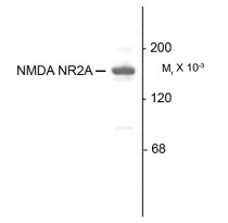
ARG52358 anti-NMDAR2A antibody WB image
Western Blot: 10 µg of Rat hippocampal (Hipp) lysate stained with ARG52358 anti-NMDAR2A antibody showing specific immunolabeling of the ~180 kDa NR2A subunit of the NMDA receptor.
-
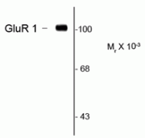
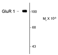
ARG52314 anti-GluR1 antibody [RH95] WB image
Western Blot: rat hippocampal lysate showing specific immunolabeling of the ~105k GluR1 protein stained with GluR1 antibody (ARG52314).
-

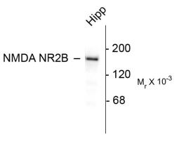
ARG52363 anti-NMDAR2B antibody WB image
Western blot: 10 ug of rat hippocampal (Hipp) lysate stained with ARG52363 anti-NMDAR2B antibody showing specific immunolabeling of the ~180k NR2B subunit of the NMDA receptor.
-

ARG52358 anti-NMDAR2A antibody IHC-Fr image
Immunohistochemistry: Rabbit retina stained with ARG52358 anti-NMDAR2A antibody showing NR2A in the rod and cone photoreceptors in the outer plexiform layer as well as the entire inner plexiform layer.
-
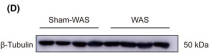
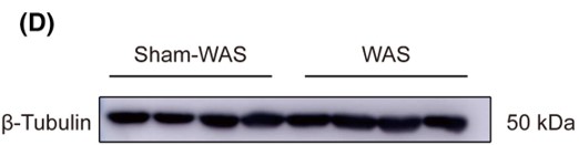
ARG65350 Goat anti-Mouse IgG antibody (HRP) WB image
Western blot: Rat basolateral amygdala stained with ARG62347 anti-beta Tubulin antibody [BT7R] at 1:1000 dilution, ARG65350 Goat anti-Mouse IgG antibody (HRP) at 1:5000 dilution.
From Guang-Bing Duan et al. CNS Neurosci Ther. (2024), doi: 10.1111/cns.14611, Fig. 4.D.
-
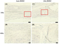
ARG65350 Goat anti-Mouse IgG antibody (HRP) IHC-P image
From Cheng-Feng Chu et al. J Pers Med. (2021), doi: 10.3390/jpm11121326, Fig. 6.
-

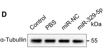
ARG65351 Goat anti-Rabbit IgG antibody (HRP) WB image
Western blot: Mouse retina stained with ARG65693 anti-alpha Tubulin antibody and ARG65351 Goat anti-Rabbit IgG antibody (HRP)
From Xiaoyuan Ye et al. Mol Ther Nucleic Acids. (2024), doi: 10.1016/j.omtn.2024.102209, Fig. 5.D.
-
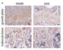
ARG65351 Goat anti-Rabbit IgG antibody (HRP) IHC-P image
From Yu-Qian Song et al. J Mol Med (Berl) (2022), doi: 10.1007/s00109-021-02165-0, Fig. 5.c.


Specific References
Expression and correlation of Surfeit 4 gene in esophageal squamous cell carcinoma
ARG65350; WB /


Environmental acidification drives inter-organ energy mobilization to enhance reproductive performance in medaka (Oryzias latipes)
ARG65350; WB /
KDF1 Promoted Proliferation, Migration and Invasion of Lung Adenocarcinoma Cells through Activating STAT3 and AKT Pathway
ARG65350: WB /


