ARG52358
anti-NMDAR2A antibody
anti-NMDAR2A antibody for IHC-Frozen sections,Western blot and Mouse,Rat,Rabbit
Neuroscience antibody; Postsynaptic Receptor antibody
Overview
| Product Description | Rabbit Polyclonal antibody recognizes NMDAR2A |
|---|---|
| Tested Reactivity | Ms, Rat, Rb |
| Predict Reactivity | Hu |
| Tested Application | IHC-Fr, WB |
| Host | Rabbit |
| Clonality | Polyclonal |
| Isotype | IgG |
| Target Name | NMDAR2A |
| Antigen Species | Rat |
| Immunogen | Fusion protein from the C-terminal region of the Rat NR2A subunit |
| Conjugation | Un-conjugated |
| Alternate Names | FESD; Glutamate receptor ionotropic, NMDA 2A; NR2A; GluN2A; N-methyl D-aspartate receptor subtype 2A; EPND; Glutamate [NMDA] receptor subunit epsilon-1; NMDAR2A; LKS; hNR2A |
Application Instructions
| Application Suggestion |
|
||||||
|---|---|---|---|---|---|---|---|
| Application Note | Specific for the ~180k NR2A subunit of the NMDA receptor. Recognizes human, mouse and Rat forms of the NR2A subunit of NMDAR. No reactivity towards the NR2B and NR2C subunits. Immunolabeling is blocked by pre-adsorption of antibody with the fusion protein used to geneRate the antibody. * The dilutions indicate recommended starting dilutions and the optimal dilutions or concentrations should be determined by the scientist. |
Properties
| Form | Liquid |
|---|---|
| Purification | Affinity purification with immunogen. |
| Buffer | 10 mM HEPES (pH 7.5), 150 mM NaCl, 100 µg BSA per ml and 50% Glycerol |
| Stabilizer | 100 µg BSA per ml, 50% Glycerol |
| Storage Instruction | For continuous use, store undiluted antibody at 2-8°C for up to a week. For long-term storage, aliquot and store at -20°C. Storage in frost free freezers is not recommended. Avoid repeated freeze/thaw cycles. Suggest spin the vial prior to opening. The antibody solution should be gently mixed before use. |
| Note | For laboratory research only, not for drug, diagnostic or other use. |
Bioinformation
| Database Links |
Swiss-port # P35436 Mouse Glutamate receptor ionotropic, NMDA 2A Swiss-port # Q00959 Rat Glutamate receptor ionotropic, NMDA 2A |
|---|---|
| Gene Symbol | GRIN2A |
| Gene Full Name | glutamate receptor, ionotropic, N-methyl D-aspartate 2A |
| Background | The ion channels activated by glutamate are typically divided into two classes. Glutamate receptors that are activated by kainate and α-amino-3-hydroxy-5-methyl-4-isoxalone propionic acid (AMPA) are known as kainate/AMPA receptors (K/AMPAR). Those that are sensitive to N-methyl-Daspartate (NMDA) are designated NMDA receptors (NMDAR). The NMDAR plays an essential role in memory, neuronal development and it has also been implicated in several disorders of the central nervous system including Alzheimer’s, epilepsy and ischemic neuronal cell death (Grosshans et al., 2002; Wenthold et al., 2003; Carroll and Zukin, 2002). The NMDA receptor is also one of the principal molecular targets for alcohol in the CNS (Lovinger et al., 1989; Alvestad et al., 2003; Snell et al., 1996). The NMDAR is also potentiated by protein phosphorylation (Lu et al., 1999). The rat NMDAR1 (NR1) was the first subunit of the NMDAR to be cloned. The NR1 protein can form NMDA activated channels when expressed in Xenopus oocytes but the currents in such channels are much smaller than those seen in situ. Channels with more physiological characteristics are produced when the NR1 subunit is combined with one or more of the NMDAR2 (NR2 A-D) subunits |
| Highlight | Related Antibody Duos and Panels: ARG30131 Postsynaptic Receptor Antibody Panel (NMDAR2A, NMDAR2B, GluR1) Related products: NMDAR2A antibodies; NMDAR2A Duos / Panels; Anti-Rabbit IgG secondary antibodies; |
| Research Area | Neuroscience antibody; Postsynaptic Receptor antibody |
| Calculated MW | 165 kDa |
Images (2) Click the Picture to Zoom In
-
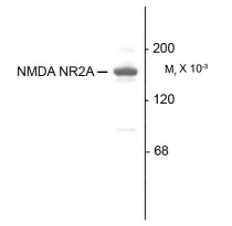
ARG52358 anti-NMDAR2A antibody WB image
Western Blot: 10 µg of Rat hippocampal (Hipp) lysate stained with ARG52358 anti-NMDAR2A antibody showing specific immunolabeling of the ~180 kDa NR2A subunit of the NMDA receptor.
-

ARG52358 anti-NMDAR2A antibody IHC-Fr image
Immunohistochemistry: Rabbit retina stained with ARG52358 anti-NMDAR2A antibody showing NR2A in the rod and cone photoreceptors in the outer plexiform layer as well as the entire inner plexiform layer.


