ARG30126
Phospho c-Myc Antibody Panel (Total, pT58, pS62)
Cancer antibody; Controls and Markers antibody; Developmental Biology antibody; Gene Regulation antibody; Immune System antibody; Signaling Transduction antibody
Component
| Cat No | Component Name | Host clonality | Reactivity | Application | Package |
|---|---|---|---|---|---|
| ARG51034 | anti-c-Myc antibody | Rabbit pAb | Hu, Ms, Rat | IHC-P, WB | 50 μl |
| ARG51538 | anti-Myc phospho (Thr58) antibody | Rabbit pAb | Hu, Ms, Rat | IHC-P, WB | 50 μl |
| ARG51785 | anti-Myc phospho (Ser62) antibody | Rabbit pAb | Hu, Ms, Rat | ICC/IF, WB | 50 μl |
| ARG65351 | Goat anti-Rabbit IgG antibody (HRP) | Goat pAb | Rb | ELISA, IHC-P, WB | 50 μl |
Overview
| Target Name | c-Myc |
|---|---|
| Alternate Names | Phospho c-Myc antibody; c-Myc antibody; Myc phospho (Thr58) antibody; Myc phospho (Ser62) antibody |
Properties
| Storage Instruction | For continuous use, store undiluted antibody at 2-8°C for up to a week. For long-term storage, aliquot and store at -20°C or below. Storage in frost free freezers is not recommended. Avoid repeated freeze/thaw cycles. Suggest spin the vial prior to opening. The antibody solution should be gently mixed before use. |
|---|---|
| Note | For laboratory research only, not for drug, diagnostic or other use. |
Bioinformation
| Gene Full Name | Antibody Panel for Phospho c-Myc (Total, pT58, pS62) |
|---|---|
| Highlight | Related Product: anti-c-Myc antibody; anti-Myc phospho (Thr58) antibody; anti-Myc phospho (Ser62) antibody; Goat anti-Rabbit IgG antibody (HRP); |
| Research Area | Cancer antibody; Controls and Markers antibody; Developmental Biology antibody; Gene Regulation antibody; Immune System antibody; Signaling Transduction antibody |
Images (11) Click the Picture to Zoom In
-
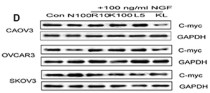
ARG51034 anti-c-Myc antibody WB image
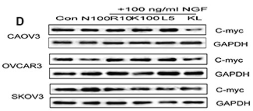
Western blot: OVCAR3 cells stained with ARG51034 anti-c-Myc antibody.
From Bo Li et al. Oncotarget. (2016), doi: 10.18632/oncotarget.13186, Fig. 8D.
-
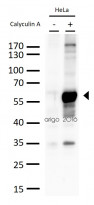
ARG51785 anti-Myc phospho (Ser62) antibody WB image
Western blot: 30 µg of HeLa cell lysates untreated or treated with calyculin A (50nM, 30mins). The blots were stained with ARG51785 anti-Myc phospho (Ser62) antibody at 1:500 dilution.
-

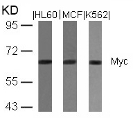
ARG51034 anti-c-Myc antibody WB image
Western Blot: extracts from HL60, MCF and K562 cells stained with anti-c-Myc antibody ARG51034.
-
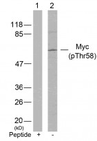
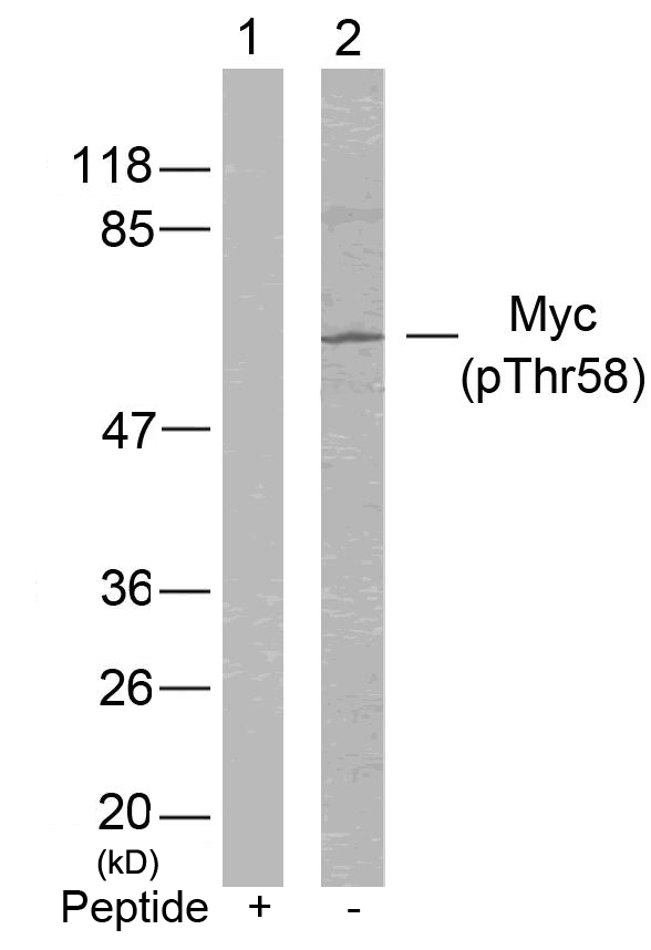
ARG51538 anti-Myc phospho (Thr58) antibody WB image
Western Blot: extracts from HeLa cells stained with anti-c-Myc (phospho Thr58) antibody ARG51538 (Lane 2) and the same antibody preincubated with blocking peptide (Lane1).
-
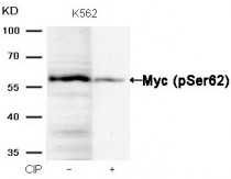
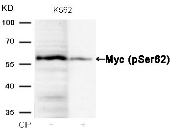
ARG51785 anti-Myc phospho (Ser62) antibody WB image
Western Blot: extracts from K562 cells, treated with calf intestinal phosphatase (CIP), stained with anti-c-Myc (phospho Ser62) antibody ARG51785.
-
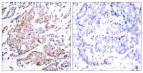
ARG51034 anti-c-Myc antibody IHC-P image
Immunohistochemistry: paraffin-embedded human breast carcinoma tissue stained with anti-c-Myc antibody ARG51034 (left) or the same antibody preincubated with blocking peptide (right).
-
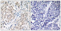
ARG51538 anti-Myc phospho (Thr58) antibody IHC-P image
Immunohistochemistry: paraffin-embedded human breast carcinoma tissue stained with anti-c-Myc (phospho Thr58) antibody ARG51538 (left) or the same antibody preincubated with blocking peptide (right).
-
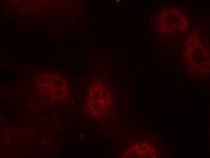
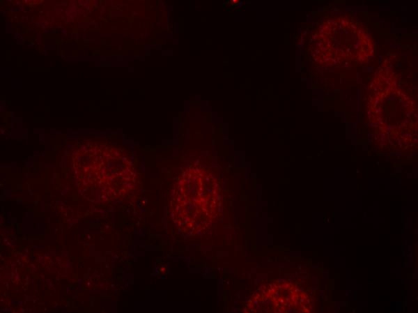
ARG51785 anti-Myc phospho (Ser62) antibody ICC/IF image
Immunofluorescence: methanol-fixed HeLa cells stained with anti-c-Myc (phospho Ser62) antibody ARG51785.
-

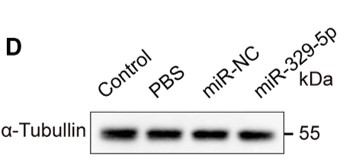
ARG65351 Goat anti-Rabbit IgG antibody (HRP) WB image
Western blot: Rat placental stained with ARG57589 anti-MTNR1A antibody at 1:1000 dilution, ARG65351 Goat anti-Rabbit IgG antibody (HRP) at 1:5000 dilution.
From Jinzhi Li et al. J Reprod Immunol. (2023), doi: 10.1016/j.jri.2023.104166, Fig. 2.B.
-

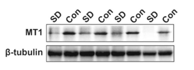
ARG65351 Goat anti-Rabbit IgG antibody (HRP) WB image
Western blot: Mouse retina stained with ARG65693 anti-alpha Tubulin antibody and ARG65351 Goat anti-Rabbit IgG antibody (HRP)
From Xiaoyuan Ye et al. Mol Ther Nucleic Acids. (2024), doi: 10.1016/j.omtn.2024.102209, Fig. 5.D.
-
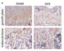
ARG65351 Goat anti-Rabbit IgG antibody (HRP) IHC-P image
From Yu-Qian Song et al. J Mol Med (Berl) (2022), doi: 10.1007/s00109-021-02165-0, Fig. 5.c.


Specific References
The Therapeutic Potential of Exosomes vs. Matrix-Bound Nanovesicles from Human Umbilical Cord Mesenchymal Stromal Cells in Osteoarthritis Treatment
ARG65351; WB /


Environmental acidification drives inter-organ energy mobilization to enhance reproductive performance in medaka (Oryzias latipes)
ARG65351; WB /
Nerve growth factor modulates the tumor cells migration in ovarian cancer through the WNT/β-catenin pathway.
ARG51034: WB / Human


