ARG51034
anti-c-Myc antibody
anti-c-Myc antibody for IHC-Formalin-fixed paraffin-embedded sections,Western blot and Human,Mouse,Rat
Cancer antibody; Controls and Markers antibody; Developmental Biology antibody; Gene Regulation antibody; Signaling Transduction antibody
Overview
| Product Description | Rabbit Polyclonal antibody recognizes c-Myc |
|---|---|
| Tested Reactivity | Hu, Ms, Rat |
| Tested Application | IHC-P, WB |
| Host | Rabbit |
| Clonality | Polyclonal |
| Isotype | IgG |
| Target Name | c-Myc |
| Antigen Species | Human |
| Immunogen | Peptide sequence around aa.356~360 (R-R-T-H-N) derived from Human Myc. |
| Conjugation | Un-conjugated |
| Alternate Names | c-Myc; MRTL; MYCC; Class E basic helix-loop-helix protein 39; Proto-oncogene c-Myc; bHLHe39; Myc proto-oncogene protein; Transcription factor p64 |
Application Instructions
| Application Suggestion |
|
||||||
|---|---|---|---|---|---|---|---|
| Application Note | * The dilutions indicate recommended starting dilutions and the optimal dilutions or concentrations should be determined by the scientist. |
Properties
| Form | Liquid |
|---|---|
| Purification | Antibodies were produced by immunizing rabbits with KLH-conjugated synthetic peptide. Antibodies were purified by affinity-chromatography using epitope-specific peptide. |
| Buffer | PBS (without Mg2+ and Ca2+, pH 7.4), 150mM NaCl, 0.02% Sodium azide and 50% Glycerol. |
| Preservative | 0.02% Sodium azide |
| Stabilizer | 50% Glycerol |
| Concentration | 1 mg/ml |
| Storage Instruction | For continuous use, store undiluted antibody at 2-8°C for up to a week. For long-term storage, aliquot and store at -20°C. Storage in frost free freezers is not recommended. Avoid repeated freeze/thaw cycles. Suggest spin the vial prior to opening. The antibody solution should be gently mixed before use. |
| Note | For laboratory research only, not for drug, diagnostic or other use. |
Bioinformation
| Database Links | |
|---|---|
| Gene Symbol | MYC |
| Gene Full Name | v-myc avian myelocytomatosis viral oncogene homolog |
| Background | Myc proto-oncogene encodes nuclear DNA-binding phosphoproteins that are involved in the regulation of gene expression and DNA replication during cell growth and differentiation. Myc encodes a protein of 65 kDa which is expressed in almost all normal and transformed cells. The expression correlates with the proliferation state of the cells. Transcription is repressed in quiescent or terminally differentiated cells. Expression of Myc is generally induced after mitogenic stimulation of cells or serum induction. Myc therefore is an important positive regulator of cell growth and proliferation. Myc has been demonstrated also to be a potent inducer of apoptosis when expressed in the absence of serum or growth factors. Apoptosis may serve also as a protective mechanism to prevent tumorigenicity elicited by deregulated Myc expression. Sequences of the Myc oncogene have been highly conserved throughout evolution, from Drosophila to vertebrates |
| Function | Transcription factor that binds DNA in a non-specific manner, yet also specifically recognizes the core sequence 5'-CAC[GA]TG-3'. Activates the transcription of growth-related genes. [UniProt] |
| Highlight | Related Antibody Duos and Panels: ARG30124 Phospho c-Myc Antibody Duo (Total, pT58) ARG30125 Phospho c-Myc Antibody Duo (Total, pS62) ARG30126 Phospho c-Myc Antibody Panel (Total, pT58, pS62) Related products: c-Myc antibodies; c-Myc Duos / Panels; Anti-Rabbit IgG secondary antibodies; |
| Research Area | Cancer antibody; Controls and Markers antibody; Developmental Biology antibody; Gene Regulation antibody; Signaling Transduction antibody |
| Calculated MW | 49 kDa |
| PTM | Phosphorylated by PRKDC. Phosphorylation at Ser-329 by PIM2 leads to the stabilization of MYC (By similarity). Phosphorylation at Ser-62 by CDK2 prevents Ras-induced senescence. Phosphorylated at Ser-62 by DYRK2; this primes the protein for subsequent phosphorylation by GSK3B at Thr-58. Phosphorylation at Thr-58 and Ser-62 by GSK3 is required for ubiquitination and degradation by the proteasome. Ubiquitinated by the SCF(FBXW7) complex when phosphorylated at Thr-58 and Ser-62, leading to its degradation by the proteasome. In the nucleoplasm, ubiquitination is counteracted by USP28, which interacts with isoform 1 of FBXW7 (FBW7alpha), leading to its deubiquitination and preventing degradation. In the nucleolus, however, ubiquitination is not counteracted by USP28, due to the lack of interaction between isoform 4 of FBXW7 (FBW7gamma) and USP28, explaining the selective MYC degradation in the nucleolus. Also polyubiquitinated by the DCX(TRUSS) complex. Ubiquitinated by TRIM6 in a phosphorylation-independent manner (By similarity). |
Images (3) Click the Picture to Zoom In
-
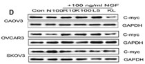
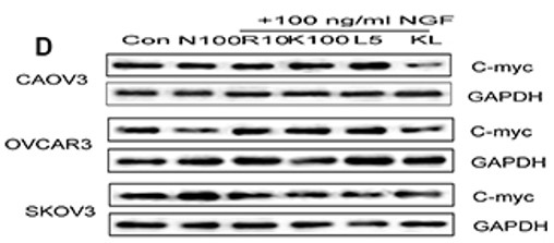
ARG51034 anti-c-Myc antibody WB image
Western blot: OVCAR3 cells stained with ARG51034 anti-c-Myc antibody.
From Bo Li et al. Oncotarget. (2016), doi: 10.18632/oncotarget.13186, Fig. 8D.
-

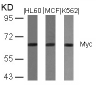
ARG51034 anti-c-Myc antibody WB image
Western Blot: extracts from HL60, MCF and K562 cells stained with anti-c-Myc antibody ARG51034.
-
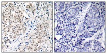
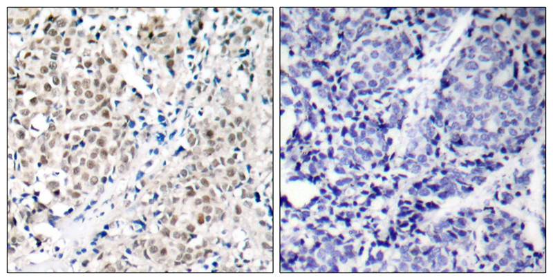
ARG51034 anti-c-Myc antibody IHC-P image
Immunohistochemistry: paraffin-embedded human breast carcinoma tissue stained with anti-c-Myc antibody ARG51034 (left) or the same antibody preincubated with blocking peptide (right).
Specific References
Nerve growth factor modulates the tumor cells migration in ovarian cancer through the WNT/β-catenin pathway.
WB / Human


