ARG30303
Nucleolar Marker Antibody Duo
Component
| Cat No | Component Name | Host clonality | Reactivity | Application | Package |
|---|---|---|---|---|---|
| ARG52280 | anti-Fibrillarin antibody [38F3] | Mouse mAb | Hu, Ms, Ce, Dm, Plnt, S. pombe | ICC/IF, IHC-Fr, WB | 50 μl |
| ARG55684 | anti-NPM1 / Nucleophosmin antibody | Rabbit pAb | Hu | FACS, ICC/IF, IHC-P, WB | 50 μl |
Overview
| Product Description | The nucleolus is a subnuclear structure with multiple roles involving ribosome biogenesis as well as cell proliferation, growth, survival, and stress response signaling. Three main components are found in the nucleolus: the granular component (GC), fibrillar center (FC), and dense fibrillar component (DFC). The GC contains nucleophosmin which is involved in ribosome biogenesis. The DFC contains the fibrillarin which is critical for rRNA processing. Accumulated evidences prove the link between nucleolar activity with cancer, virus replication, and stress. arigo's Nucleolar Marker Antibody Duo comprises GC marker nucleophosmin antibody and DFC marker fibrillarin antibody. It is an excellent solution for investigating the role of nucleolus in gene expression and stress response. |
|---|---|
| Target Name | Nucleolar Marker |
| Alternate Names | Nucleolar Marker antibody; Fibrillarin antibody; NPM1 / Nucleophosmin antibody |
Properties
| Storage Instruction | For continuous use, store undiluted antibody at 2-8°C for up to a week. For long-term storage, aliquot and store at -20°C or below. Storage in frost free freezers is not recommended. Avoid repeated freeze/thaw cycles. Suggest spin the vial prior to opening. The antibody solution should be gently mixed before use. |
|---|---|
| Note | For laboratory research only, not for drug, diagnostic or other use. |
Bioinformation
| Gene Full Name | Antibody Duo for Nucleolar Marker |
|---|---|
| Highlight | Related Product: anti-Fibrillarin antibody; anti-NPM1 / Nucleophosmin antibody; |
Images (8) Click the Picture to Zoom In
-
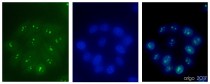
ARG52280 anti-Fibrillarin antibody [38F3] ICC/IF image
Immunofluorescence: 100% Methanol fixed (RT, 10 min) HeLa cells stained with ARG52280 anti-Fibrillarin antibody [38F3] at 1:500 dilution. Left: primary antibody (green). Middle: DAPI (blue). Right: Merge.
Secondary antibody: ARG55393 Goat anti-Mouse IgG (H+L) antibody (FITC)
-
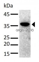
ARG52280 anti-Fibrillarin antibody [38F3] WB image
Western blot: 30 µg of HeLa cell lysate stained with ARG52280 anti-Fibrillarin antibody [38F3] at 1:1000 dilution.
-
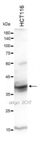
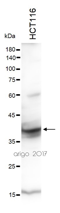
ARG55684 anti-NPM1 / Nucleophosmin antibody WB image
Western blot: 30 µg of HCT116 cell lysate stained with ARG55684 anti-NPM1 / Nucleophosmin antibody at 1:1000 dilution.
-
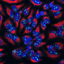
ARG52280 anti-Fibrillarin antibody [38F3] ICC/IF image
Immunofluorescence: HeLa cells stained with ARG52280 anti-Fibrillarin antibody [38F3] (green) at 1:100 dilution, and costained with ARG52468 anti-Vimentin antibody (red) at 1:1000 dilution. DAPI (blue) for nuclear staining.
The Fibrillarin antibody shows strong staining of nucleoli in the nucleus, while the Vimentin antibody reveals cytoplasmic intermediate filaments.
-

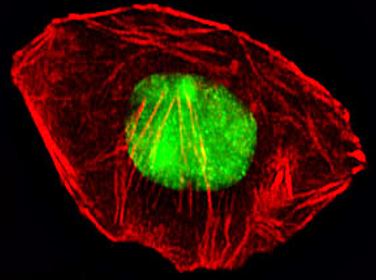
ARG55684 anti-NPM1 / Nucleophosmin antibody ICC/IF image
Immunofluorescence: A549 cells were fixed with 4% PFA (20 min), permeabilized with Triton X-100 (0.1%, 10 min), then stained with ARG55684 anti-NPM1 / Nucleophosmin antibody (green) at 1:25 dilution, 1 hour at 37°C. Cytoplasmic actin was counterstained with Alexa Fluor® 555 (red) conjugated Phalloidin (7 units/ml, 1 hour at 37°C).
-
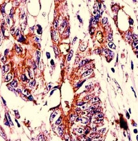
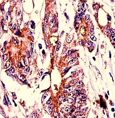
ARG55684 anti-NPM1 / Nucleophosmin antibody IHC-P image
Immunohistochemistry: Formalin-fixed and paraffin-embedded Human colon carcinoma stained with ARG55684 anti-NPM1 / Nucleophosmin antibody.
-
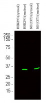
ARG52280 anti-Fibrillarin antibody [38F3] WB image
Western blot: HEK293 cytosol, HEK293 nuclear, NIH/3T3 cytosol and NIH/3T3 nuclear fractions stained with ARG52280 anti-Fibrillarin antibody [38F3] (green) at 1:500 dilution.
-
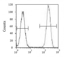
ARG55684 anti-NPM1 / Nucleophosmin antibody FACS image
Flow Cytometry: HeLa cells stained with ARG55684 anti-NPM1 / Nucleophosmin antibody (right histogram) or without primary antibody as control (left histogram), followed by incubation with FITC labelled secondary antibody.