ARG52280
anti-Fibrillarin antibody [38F3]
anti-Fibrillarin antibody [38F3] for ICC/IF,IHC-Frozen sections,Western blot and Human,Mouse,C. elegans,D. melanogaster ,Plant,S. pombe
Gene Regulation antibody; Nucleolar Marker antibody; DFC Marker antibody; Dense fibrillar component Marker antibody

Overview
| Product Description | Mouse Monoclonal antibody [38F3] recognizes Fibrillarin |
|---|---|
| Tested Reactivity | Hu, Ms, Ce, Dm, Plnt, S. pombe |
| Tested Application | ICC/IF, IHC-Fr, WB |
| Host | Mouse |
| Clonality | Monoclonal |
| Clone | 38F3 |
| Isotype | IgG1 |
| Target Name | Fibrillarin |
| Antigen Species | Yeast |
| Immunogen | Yeast nuclear preparations |
| Epitope | EYRAWNPFRSKLAAAILGGV |
| Conjugation | Un-conjugated |
| Alternate Names | rRNA 2'-O-methyltransferase fibrillarin; RNU3IP1; 34 kDa nucleolar scleroderma antigen; FIB; FLRN; EC 2.1.1.-; Histone-glutamine methyltransferase |
Application Instructions
| Application Suggestion |
|
||||||||
|---|---|---|---|---|---|---|---|---|---|
| Application Note | Specific for the ~34kDa Fibrillarin /Nop1p protein. * The dilutions indicate recommended starting dilutions and the optimal dilutions or concentrations should be determined by the scientist. |
Properties
| Form | Liquid |
|---|---|
| Purification | Total IgG fraction |
| Buffer | Total IgG fraction and 10 mM Sodium azide |
| Preservative | 10 mM Sodium azide |
| Storage Instruction | For continuous use, store undiluted antibody at 2-8°C for up to a week. For long-term storage, aliquot and store at -20°C or below. Storage in frost free freezers is not recommended. Avoid repeated freeze/thaw cycles. Suggest spin the vial prior to opening. The antibody solution should be gently mixed before use. |
| Note | For laboratory research only, not for drug, diagnostic or other use. |
Bioinformation
| Database Links |
Swiss-port # P22087 Human rRNA 2'-O-methyltransferase fibrillarin Swiss-port # P35550 Mouse rRNA 2'-O-methyltransferase fibrillarin |
|---|---|
| Gene Symbol | NOP1 |
| Background | Nop1p was originally identified as a nucleolar protein of bakers yeast, Saccharomyces cerevisiae. The Nop1p protein is 327 amino acids in size (34.5kDa), is essential for yeast viability, and is localized in the nucleoli . The systematic name for S. cerevisiae Nop1 is YDL014W, and it is now known to be part of the small subunit processome complex, involved in the processing of pre-18S ribosomal RNA. Nop1p is the yeast homologue of a protein found in all eukaryotes and archea generally called fibrillarin . Fibrillarin/Nop1p is extraordinarily conserved, so that the yeast and human proteins are 67% identical, and the human protein can functionally replace the yeast protein. Patients with the autoimmune disease scleroderma often have strong circulating autoantibodies to a ~34kDa protein which was subsequently found to be fibrillarin. Recent studies show that knock-out of the fibrillarin gene in mice results in embryonic lethality, although mice with only one functional fibrillarin/Nop1p gene were viable . This antibody is becoming widely used as a convenient marker for nucleoli in a wide variety of species (e.g. 4-6). |
| Highlight | Related Antibody Duos and Panels: ARG30303 Nucleolar Marker Antibody Duo Related products: Fibrillarin antibodies; Fibrillarin Duos / Panels; Anti-Mouse IgG secondary antibodies; Related poster download: Organelle Markers & Loading Control |
| Research Area | Gene Regulation antibody; Nucleolar Marker antibody; DFC Marker antibody; Dense fibrillar component Marker antibody |
| Calculated MW | 34 kDa |
| PTM | By homology to other fibrillarins, some or all of the N-terminal domain arginines are modified to asymmetric dimethylarginine (DMA). |
Images (4) Click the Picture to Zoom In
-
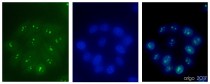
ARG52280 anti-Fibrillarin antibody [38F3] ICC/IF image
Immunofluorescence: 100% Methanol fixed (RT, 10 min) HeLa cells stained with ARG52280 anti-Fibrillarin antibody [38F3] at 1:500 dilution. Left: primary antibody (green). Middle: DAPI (blue). Right: Merge.
Secondary antibody: ARG55393 Goat anti-Mouse IgG (H+L) antibody (FITC)
-
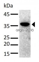
ARG52280 anti-Fibrillarin antibody [38F3] WB image
Western blot: 30 µg of HeLa cell lysate stained with ARG52280 anti-Fibrillarin antibody [38F3] at 1:1000 dilution.
-
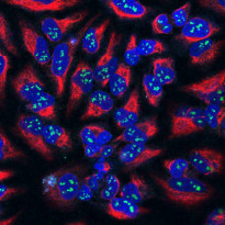
ARG52280 anti-Fibrillarin antibody [38F3] ICC/IF image
Immunofluorescence: HeLa cells stained with ARG52280 anti-Fibrillarin antibody [38F3] (green) at 1:100 dilution, and costained with ARG52468 anti-Vimentin antibody (red) at 1:1000 dilution. DAPI (blue) for nuclear staining.
The Fibrillarin antibody shows strong staining of nucleoli in the nucleus, while the Vimentin antibody reveals cytoplasmic intermediate filaments.
-
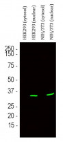
ARG52280 anti-Fibrillarin antibody [38F3] WB image
Western blot: HEK293 cytosol, HEK293 nuclear, NIH/3T3 cytosol and NIH/3T3 nuclear fractions stained with ARG52280 anti-Fibrillarin antibody [38F3] (green) at 1:500 dilution.


Customer's Feedback
Excellent

Review for anti-Fibrillarin antibody [38F3]
Application:IF/ICC
Sample:HeLa
Fixation Buffer:100% Methanol
Fixation Time:10 min
Fixation Temperature:RT ºC
Permeabilization Buffer:0.1% Triton X-100
Primary Antibody Dilution Factor:1:500
Primary Antibody Incubation Time:overnight
Primary Antibody Incubation Temperature:4 ºC
Conjugation of Secondary Antibody:FITC
Excellent

Review for anti-Fibrillarin antibody [38F3]
Application:WB
Sample:HeLa
Sample Loading Amount:30 µg
Primary Antibody Dilution Factor:1:1000
Primary Antibody Incubation Time:overnight
Primary Antibody Incubation Temperature:4 ºC
Clone References
A novel mechanism inducing genome instability in Kaposi's sarcoma-associated herpesvirus infected cells.
WB /


The transcription factor EGR1 localizes to the nucleolus and is linked to suppression of ribosomal precursor synthesis.
ICC/IF / Human
Def functions as a cell autonomous factor in organogenesis of digestive organs in zebrafish.
IHC-Fr /
Involvement of phosphatidylinositol 4,5-bisphosphate in RNA polymerase I transcription.
ICC/IF / Human
Arabidopsis DEAD-box RNA helicase UAP56 interacts with both RNA and DNA as well as with mRNA export factors.
ICC/IF / Arabidopsis thaliana
Developmental regulation of nucleolus size during Drosophila eye differentiation.
IHC / Fruit fly (Drosophila melanogaster)
40S ribosome biogenesis co-factors are essential for gametophyte and embryo development.
IHC / Plant
MicroRNA miR-308 regulates dMyc through a negative feedback loop in Drosophila.
WB / Fruit fly (Drosophila melanogaster)


