ARG55684
anti-NPM1 / Nucleophosmin antibody
anti-NPM1 / Nucleophosmin antibody for Flow cytometry,ICC/IF,IHC-Formalin-fixed paraffin-embedded sections,Western blot and Human
Nucleolar Marker antibody; GC Marker antibody; Granular Component Marker antibody

Overview
| Product Description | Rabbit Polyclonal antibody recognizes NPM1 / Nucleophosmin |
|---|---|
| Tested Reactivity | Hu |
| Tested Application | FACS, ICC/IF, IHC-P, WB |
| Host | Rabbit |
| Clonality | Polyclonal |
| Isotype | IgG |
| Target Name | NPM1 / Nucleophosmin |
| Antigen Species | Human |
| Immunogen | KLH-conjugated synthetic peptide corresponding to aa. 198-226 (C-terminus) of Human NPM1. |
| Conjugation | Un-conjugated |
| Alternate Names | NPM; Nucleolar protein NO38; B23; Nucleophosmin; Numatrin; Nucleolar phosphoprotein B23 |
Application Instructions
| Application Suggestion |
|
||||||||||
|---|---|---|---|---|---|---|---|---|---|---|---|
| Application Note | * The dilutions indicate recommended starting dilutions and the optimal dilutions or concentrations should be determined by the scientist. |
Properties
| Form | Liquid |
|---|---|
| Purification | This antibody is prepared by Saturated Ammonium Sulfate (SAS) precipitation followed by dialysis against PBS. |
| Buffer | PBS and 0.09% (W/V) Sodium azide. |
| Preservative | 0.09% (W/V) Sodium azide. |
| Storage Instruction | For continuous use, store undiluted antibody at 2-8°C for up to a week. For long-term storage, aliquot and store at -20°C or below. Storage in frost free freezers is not recommended. Avoid repeated freeze/thaw cycles. Suggest spin the vial prior to opening. The antibody solution should be gently mixed before use. |
| Note | For laboratory research only, not for drug, diagnostic or other use. |
Bioinformation
| Database Links | |
|---|---|
| Gene Symbol | NPM1 |
| Gene Full Name | nucleophosmin (nucleolar phosphoprotein B23, numatrin) |
| Background | This gene encodes a phosphoprotein which moves between the nucleus and the cytoplasm. The gene product is thought to be involved in several processes including regulation of the ARF/p53 pathway. A number of genes are fusion partners have been characterized, in particular the anaplastic lymphoma kinase gene on chromosome 2. Mutations in this gene are associated with acute myeloid leukemia. More than a dozen pseudogenes of this gene have been identified. Alternative splicing results in multiple transcript variants.[provided by RefSeq, Nov 2009] |
| Function | Involved in diverse cellular processes such as ribosome biogenesis, centrosome duplication, protein chaperoning, histone assembly, cell proliferation, and regulation of tumor suppressors p53/TP53 and ARF. Binds ribosome presumably to drive ribosome nuclear export. Associated with nucleolar ribonucleoprotein structures and bind single-stranded nucleic acids. Acts as a chaperonin for the core histones H3, H2B and H4. Stimulates APEX1 endonuclease activity on apurinic/apyrimidinic (AP) double-stranded DNA but inhibits APEX1 endonuclease activity on AP single-stranded RNA. May exert a control of APEX1 endonuclease activity within nucleoli devoted to repair AP on rDNA and the removal of oxidized rRNA molecules. In concert with BRCA2, regulates centrosome duplication. Regulates centriole duplication: phosphorylation by PLK2 is able to trigger centriole replication. Negatively regulates the activation of EIF2AK2/PKR and suppresses apoptosis through inhibition of EIF2AK2/PKR autophosphorylation. [UniProt] |
| Cellular Localization | Cytoplasm, Cytoskeleton, Nucleus |
| Highlight | Related Antibody Duos and Panels: ARG30303 Nucleolar Marker Antibody Duo Related products: NPM1 antibodies; NPM1 Duos / Panels; Anti-Rabbit IgG secondary antibodies; |
| Research Area | Nucleolar Marker antibody; GC Marker antibody; Granular Component Marker antibody |
| Calculated MW | 33 kDa |
| PTM | Acetylated at C-terminal lysine residues, thereby increasing affinity to histones. ADP-ribosylated. Phosphorylated at Ser-4 by PLK1 and PLK2. Phosphorylation at Ser-4 by PLK2 in S phase is required for centriole duplication and is sufficient to trigger centriole replication. Phosphorylation at Ser-4 by PLK1 takes place during mitosis. Phosphorylated by CDK2 at Ser-125 and Thr-199. Phosphorylation at Thr-199 may trigger initiation of centrosome duplication. Phosphorylated by CDK1 at Thr-199, Thr-219, Thr-234 and Thr-237 during cell mitosis. When these four sites are phosphorated, RNA-binding activity seem to be abolished. May be phosphorylated at Ser-70 by NEK2. The Thr-199 phosphorylated form has higher affinity for ROCK2. CDK6 triggers Thr-199 phosphorylation when complexed to Kaposi's sarcoma herpesvirus (KSHV) V-cyclin, leading to viral reactivation by reducing viral LANA levels. Sumoylated by ARF. May be ubiquitinated. Ubiquitination leads to proteasomal degradation. |
Images (4) Click the Picture to Zoom In
-
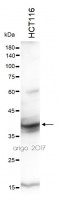
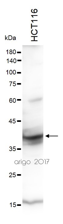
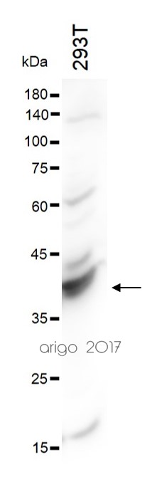
ARG55684 anti-NPM1 / Nucleophosmin antibody WB image
Western blot: 30 µg of HCT116 cell lysate stained with ARG55684 anti-NPM1 / Nucleophosmin antibody at 1:1000 dilution.
-

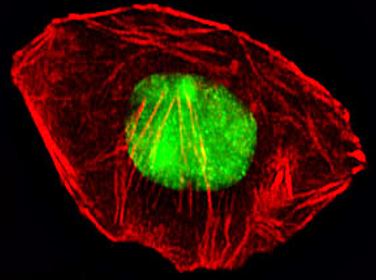
ARG55684 anti-NPM1 / Nucleophosmin antibody ICC/IF image
Immunofluorescence: A549 cells were fixed with 4% PFA (20 min), permeabilized with Triton X-100 (0.1%, 10 min), then stained with ARG55684 anti-NPM1 / Nucleophosmin antibody (green) at 1:25 dilution, 1 hour at 37°C. Cytoplasmic actin was counterstained with Alexa Fluor® 555 (red) conjugated Phalloidin (7 units/ml, 1 hour at 37°C).
-
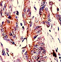
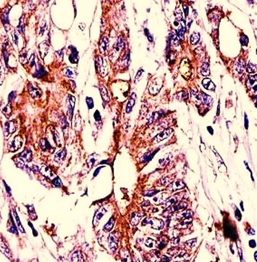
ARG55684 anti-NPM1 / Nucleophosmin antibody IHC-P image
Immunohistochemistry: Formalin-fixed and paraffin-embedded Human colon carcinoma stained with ARG55684 anti-NPM1 / Nucleophosmin antibody.
-
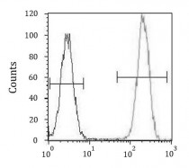
ARG55684 anti-NPM1 / Nucleophosmin antibody FACS image
Flow Cytometry: HeLa cells stained with ARG55684 anti-NPM1 / Nucleophosmin antibody (right histogram) or without primary antibody as control (left histogram), followed by incubation with FITC labelled secondary antibody.

Customer's Feedback
Good
Review for anti-NPM1 / Nucleophosmin antibody
Application:WB
Sample:293T
Sample Loading Amount:30 µg
Primary Antibody Dilution Factor:1:1000
Primary Antibody Incubation Time:overnight
Primary Antibody Incubation Temperature:4 ºC


