ARG30346
Myofibroblast / Fibrosis Antibody Panel
Component
| Cat No | Component Name | Host clonality | Reactivity | Application | Package |
|---|---|---|---|---|---|
| ARG66381 | anti-alpha smooth muscle Actin antibody [SQab18108] | Rabbit mAb | Hu, Ms, Rat, AGMK, Bov, Ctl, Dog, Pig, Zfsh | FACS, ICC/IF, IHC-Fr, IHC-P, WB | 20 μl |
| ARG21965 | anti-Collagen I antibody, pre-adsorbed | Goat pAb | Bov, Cat, Chk, Dog, Elp, Gpig, Hm, Hu, Ms, Pig, Rb, Rat, Sheep | EM, ELISA, FLISA, FACS, ICC/IF, IHC-P, IHC-Fr, IP, WB | 20 μg |
| ARG66162 | anti-Fibronectin antibody | Mouse mAb | Hu, Ms, Rat | IHC-P, WB | 20 μg |
Overview
| Product Description | Myofibroblasts are a differentiated cell type with a phenotype between a fibroblast and a smooth muscle cell. They play a physiological role in wound healing and fibrosis. Myofibroblast/Fibrosis Antibody Panel is an all-in-one solution to make the research of myofibroblast and fibrosis easy and economical. This antibody panel comprises the myofibroblast markers including smooth muscle actin, collagen I, and fibronectin antibodies. It is the best solution to study myofibroblast and fibrosis in samples from human, mouse or rat. Related news: New antibody panels for Myofibroblasts and CAFs |
|---|---|
| Target Name | Myofibroblast / Fibrosis |
Properties
| Storage Instruction | For continuous use, store undiluted antibody at 2-8°C for up to a week. For long-term storage, aliquot and store at -20°C or below. Storage in frost free freezers is not recommended. Avoid repeated freeze/thaw cycles. Suggest spin the vial prior to opening. The antibody solution should be gently mixed before use. |
|---|---|
| Note | For laboratory research only, not for drug, diagnostic or other use. |
Bioinformation
| Highlight | Related Product: anti-alpha smooth muscle Actin antibody; anti-Collagen I antibody; anti-Fibronectin antibody; |
|---|
Images (24) Click the Picture to Zoom In
-
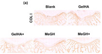
ARG21965 anti-Collagen I antibody, pre-adsorbed IHC-P image
Immunohistochemistry: Rabbit osteochondral stained with ARG21965 anti-Collagen I antibody, pre-adsorbed at 1:100 dilution.
From Wenli Dai et al. Enhanced osteochondral repair with hyaline cartilage formation using an extracellular matrix-inspired natural scaffold (2023), doi: 10.1016/j.scib.2023.07.050, Fig. 7a.
-
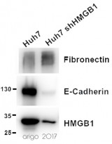
ARG66162 anti-Fibronectin antibody WB image
Western blot: 20 µg of Huh7 and Huh7 shHMGB1 cell lysates stained with ARG66162 anti-Fibronectin antibody (1:1000), ARG55914 anti-E-cadherin antibody (1:1000) and ARG65636 anti-HMGB1 antibody (1:2000).
-
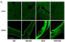
ARG66381 anti-alpha smooth muscle Actin antibody [SQab18108] IHC-Fr image
Immunohistochemistry: Mouse liver and Mouse aorta stained with ARG66381 anti-alpha smooth muscle Actin antibody [SQab18108] at 1:100 dilution.
From Masahiro Terasawa et al. Cells (2023), doi: 10.3390/cells12222666, Fig. 2A.
-
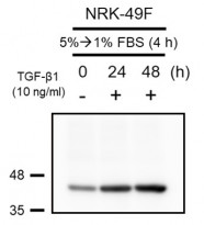
ARG66381 anti-alpha smooth muscle Actin antibody [SQab18108] WB image
Western blot: 30 μg of NRK-49F cells treated with TGF beta 1 (10 ng/ml) for 0~48 hours. Cell lysates were stained with ARG66381 anti-alpha smooth muscle Actin antibody [SQab18108] at 1:2000 dilution, overnight at 4°C.
-
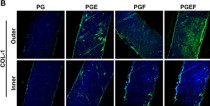
ARG21965 anti-Collagen I antibody, pre-adsorbed ICC/IF image
Immunofluorescence: Rabbit synovium-derived mesenchymal stem cell stained with ARG21965 anti-Collagen I antibody, pre-adsorbed at 1:1000 dilution.
From Zong Li e et al. Chemical Engineering Journal, (2023), doi: 10.1016/j.cej.2023.145209, Fig. 4B.
-
ARG21965 anti-Collagen I antibody, pre-adsorbed IHC-P image
Immunohistochemistry: Canine aorta mesothelial stained with ARG21965 anti-Collagen I antibody, pre-adsorbed at 1:500 dilution.
From Masakazu Shimada et al. PLoS One. (2022), doi: 35061786, Fig. 1.
-
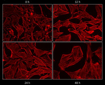
ARG66381 anti-alpha smooth muscle Actin antibody [SQab18108] ICC/IF image
Immunofluorescence: NRK-49F cells treated with TGF beta 1 (10 ng/ml) for 0~48 hours. Cells were fixed with 4% PFA for 15 min at room temperature and permeabilizated by 0.5% Triton X-100. Cells were stained with ARG66381 anti-alpha smooth muscle Actin antibody [SQab18108] at 1:200 dilution, overnight at 4°C.
-
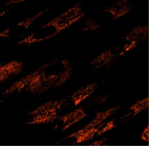
ARG21965 anti-Collagen I antibody, pre-adsorbed ICC/IF image
Immunofluorescence: Human fibroblasts were stained with ARG21965 anti-Collagen I antibody (pre-adsorbed).
-
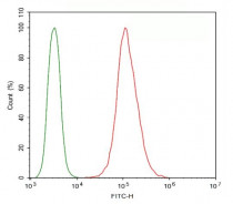
ARG66381 anti-alpha smooth muscle Actin antibody [SQab18108] FACS image
Flow Cytometry: HeLa cells were fixed with 4% paraformaldehyde (10 min) and then permeabilized with 0.1% TritonX-100 for 15 min. The cells were then stained with ARG66381 anti-alpha smooth muscle Actin antibody [SQab18108] (red) at 1:100 dilution in 1x PBS/1% BSA for 30 min at 4°C, followed by Alexa Fluor® 488 labelled secondary antibody. Unlabelled sample (green) was used as a control.
-
ARG21965 anti-Collagen I antibody, pre-adsorbed IHC-Fr image
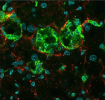
Immunohistochemistry: Frozen MC4R-KO Mouse liver section was stained with ARG21965 anti-Collagen I antibody (pre-adsorbed), anti-F4/80 antibody followed by secondary antibodies and DAPI.
-
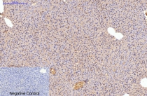
ARG66162 anti-Fibronectin antibody IHC-P image
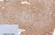
Immunohistochemistry: Paraffin-embedded Rat liver tissue stained with ARG66162 anti-Fibronectin antibody at 1:200 (4°C, overnight). Antigen Retrieval: Boil tissue section in Sodium citrate buffer (pH 6.0) for 20 min. Secondary antibody was diluted at 1:200 (RT, 30min). Negative control: Secondary antibody only.
-
ARG66381 anti-alpha smooth muscle Actin antibody [SQab18108] IHC-P image
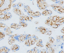
Immunohistochemistry: Formalin-fixed and paraffin-embedded placenta stained with ARG66381 anti-alpha smooth muscle Actin antibody [SQab18108] at 1:2000 dilution. Antigen Retrieval: Heat mediated was performed using Tris/EDTA buffer (pH 9.0).
-
ARG21965 anti-Collagen I antibody, pre-adsorbed WB image
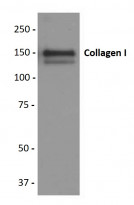
Western blot: Purified Human Type I Collagen stained with ARG21965 anti-Collagen I antibody (pre-adsorbed).
-
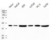
ARG66381 anti-alpha smooth muscle Actin antibody [SQab18108] WB image
Western blot: 10 µg of HeLa, HaCat, 293, LnCap, PC-3 and T47D cell lysates stained with ARG66381 anti-alpha smooth muscle Actin antibody [SQab18108] at 1:2000 dilution.
-
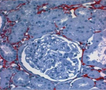
ARG21965 anti-Collagen I antibody, pre-adsorbed IHC-P image
Immunohistochemistry: Paraffin embedded Rat kidney section post uninephrectomy was stained with ARG21965 anti-Collagen I antibody (pre-adsorbed) followed by a secondary antibody and AEC.
-
ARG66162 anti-Fibronectin antibody IHC-P image
Immunohistochemistry: Paraffin-embedded Mouse liver tissue stained with ARG66162 anti-Fibronectin antibody at 1:200 (4°C, overnight). Antigen Retrieval: Boil tissue section in Sodium citrate buffer (pH 6.0) for 20 min. Secondary antibody was diluted at 1:200 (RT, 30min). Negative control: Secondary antibody only.
-
ARG66162 anti-Fibronectin antibody IHC image
Immunohistochemistry: Human appendix tissue stained with ARG66162 anti-Fibronectin antibody (red) at 1:200 (4°C, overnight). Picture A: Target. Picture B: DAPI. Picture C: merge of A and B.
-
ARG66162 anti-Fibronectin antibody IHC image
Immunohistochemistry: Human appendix tissue stained with ARG66162 anti-Fibronectin antibody (red) at 1:200 (4°C, overnight). Picture A: Target. Picture B: DAPI. Picture C: merge of A and B.
-
ARG66162 anti-Fibronectin antibody IHC image
Immunohistochemistry: Human appendix tissue stained with ARG66162 anti-Fibronectin antibody (red) at 1:200 (4°C, overnight). Picture A: Target. Picture B: DAPI. Picture C: merge of A and B.
-
ARG66162 anti-Fibronectin antibody IHC image
Immunohistochemistry: Mouse spleen tissue stained with ARG66162 anti-Fibronectin antibody (red) at 1:200 (4°C, overnight). Picture A: Target. Picture B: DAPI. Picture C: merge of A and B.
-
ARG66162 anti-Fibronectin antibody IHC image
Immunohistochemistry: Mouse spleen tissue stained with ARG66162 anti-Fibronectin antibody (red) at 1:200 (4°C, overnight). Picture A: Target. Picture B: DAPI. Picture C: merge of A and B.
-
ARG66162 anti-Fibronectin antibody IHC image
Immunohistochemistry: Mouse spleen tissue stained with ARG66162 anti-Fibronectin antibody (red) at 1:200 (4°C, overnight). Picture A: Target. Picture B: DAPI. Picture C: merge of A and B.
-
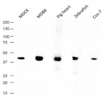
ARG66381 anti-alpha smooth muscle Actin antibody [SQab18108] WB image
Western blot: 10 µg of MDCK, MDBK, Pig heart, Zebrafish and Cos-7 lysates stained with ARG66381 anti-alpha smooth muscle Actin antibody [SQab18108] at 1:2000 dilution.
-
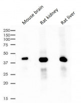
ARG66381 anti-alpha smooth muscle Actin antibody [SQab18108] WB image
Western blot: 10 µg of Mouse brain, Rat kidney and Rat liver lysates stained with ARG66381 anti-alpha smooth muscle Actin antibody [SQab18108] at 1:2000 dilution.
Specific References
