ARG21965
anti-Collagen I antibody, pre-adsorbed
anti-Collagen I antibody, pre-adsorbed for Electron microscopy,ELISA,FLISA,Flow cytometry,ICC/IF,IHC-Formalin-fixed paraffin-embedded sections,IHC-Frozen sections,Immunoprecipitation,Western blot and Bovine,Cat,Chicken,Dog,Elephant,Guinea pig,Hamster,Human,Mouse,Pig,Rabbit,Rat,Sheep
Overview
| Product Description | Goat Polyclonal antibody recognizes Collagen I |
|---|---|
| Tested Reactivity | Hu, Ms, Rat, Bov, Cat, Chk, Dog, Elp, Gpig, Hm, Pig, Rb, Sheep |
| Tested Application | ELISA, EM, FACS, FLISA, ICC/IF, IHC-Fr, IHC-P, IP, WB |
| Specificity | The antibody reacts with conformational determinants on type I collagen. The antibody is pre-adsorbed with Collagen types II, III, IV, V and VI, so the antibody may not react with Collagen types II, III, IV, V and VI. |
| Host | Goat |
| Clonality | Polyclonal |
| Isotype | IgG |
| Target Name | Collagen I |
| Antigen Species | Human |
| Immunogen | Human Type I Collagen |
| Conjugation | Un-conjugated |
| Alternate Names | collagen type I alpha 1 chain; OI1; OI2; OI3; OI4; EDSC; Collagen alpha-1(I) chain; Alpha-1 type I collagen; Collagen type I alpha 2 chain; Collagen alpha-2(I) chain |
Application Instructions
| Pre Adsorbed | Collagen types II, III, IV, V and VI. | ||||||||||||||||||||
|---|---|---|---|---|---|---|---|---|---|---|---|---|---|---|---|---|---|---|---|---|---|
| Application Suggestion |
|
||||||||||||||||||||
| Application Note | WB: The antibody reacts with native and denature form of Collagen I protein. IHC-P: Antigen retrieval: select one of below: 1. Digestion with 0.1% pepsin from porcine gastric mucosa 3200–4500 units per mg (Sigma, Vienna, Austria) in 0.5 M acetic acid for 2 h at 37°C. 2. Microwave for 4 X 5 min at 800W in 0.01 M Sodium citrate buffer (pH 6.0). * The dilutions indicate recommended starting dilutions and the optimal dilutions or concentrations should be determined by the scientist. |
||||||||||||||||||||
| Observed Size | ~120-190 kDa (depending on the sample types) |
Properties
| Form | Liquid |
|---|---|
| Purification | Affinity purification with immunogen. |
| Buffer | BBS (pH 8.2) |
| Concentration | 0.4 mg/ml |
| Storage Instruction | For continuous use, store undiluted antibody at 2-8°C for up to a week. For long-term storage, aliquot and store at -20°C. Storage in frost free freezers is not recommended. Avoid repeated freeze/thaw cycles. Suggest spin the vial prior to opening. The antibody solution should be gently mixed before use. |
| Note | For laboratory research only, not for drug, diagnostic or other use. |
Bioinformation
| Gene Symbol | COL1A1; COL1A2 |
|---|---|
| Gene Full Name | collagen, type I |
| Background | Collagen type I is a fibril-forming collagen found in most connective tissues and is abundant in bone, cornea, dermis and tendon. Mutations in this gene are associated with osteogenesis imperfecta types I-IV, Ehlers-Danlos syndrome type VIIA, Ehlers-Danlos syndrome Classical type, Caffey Disease and idiopathic osteoporosis. Reciprocal translocations between chromosomes 17 and 22, where this gene and the gene for platelet-derived growth factor beta are located, are associated with a particular type of skin tumor called dermatofibrosarcoma protuberans, resulting from unregulated expression of the growth factor. Two transcripts, resulting from the use of alternate polyadenylation signals, have been identified for this gene. [provided by R. Dalgleish, Feb 2008] |
| Function | Type I collagen is a member of group I collagen (fibrillar forming collagen). [UniProt] |
| Highlight | Related Antibody Duos and Panels: ARG30346 Myofibroblast / Fibrosis Antibody Panel Related products: Collagen I antibodies; Collagen I ELISA Kits; Collagen I Duos / Panels; Anti-Goat IgG secondary antibodies; Related news: Collagen I antibody for studying rabbit bone differentiation New antibody panels for Myofibroblasts and CAFs |
| Calculated MW | COL1A1: 139 kDa COL1A2: 129 kDa |
| PTM | Proline residues at the third position of the tripeptide repeating unit (G-X-P) are hydroxylated in some or all of the chains. Proline residues at the second position of the tripeptide repeating unit (G-P-X) are hydroxylated in some of the chains. O-linked glycan consists of a Glc-Gal disaccharide bound to the oxygen atom of a post-translationally added hydroxyl group. |
Images (7) Click the Picture to Zoom In
-
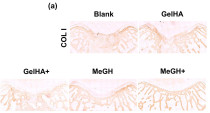
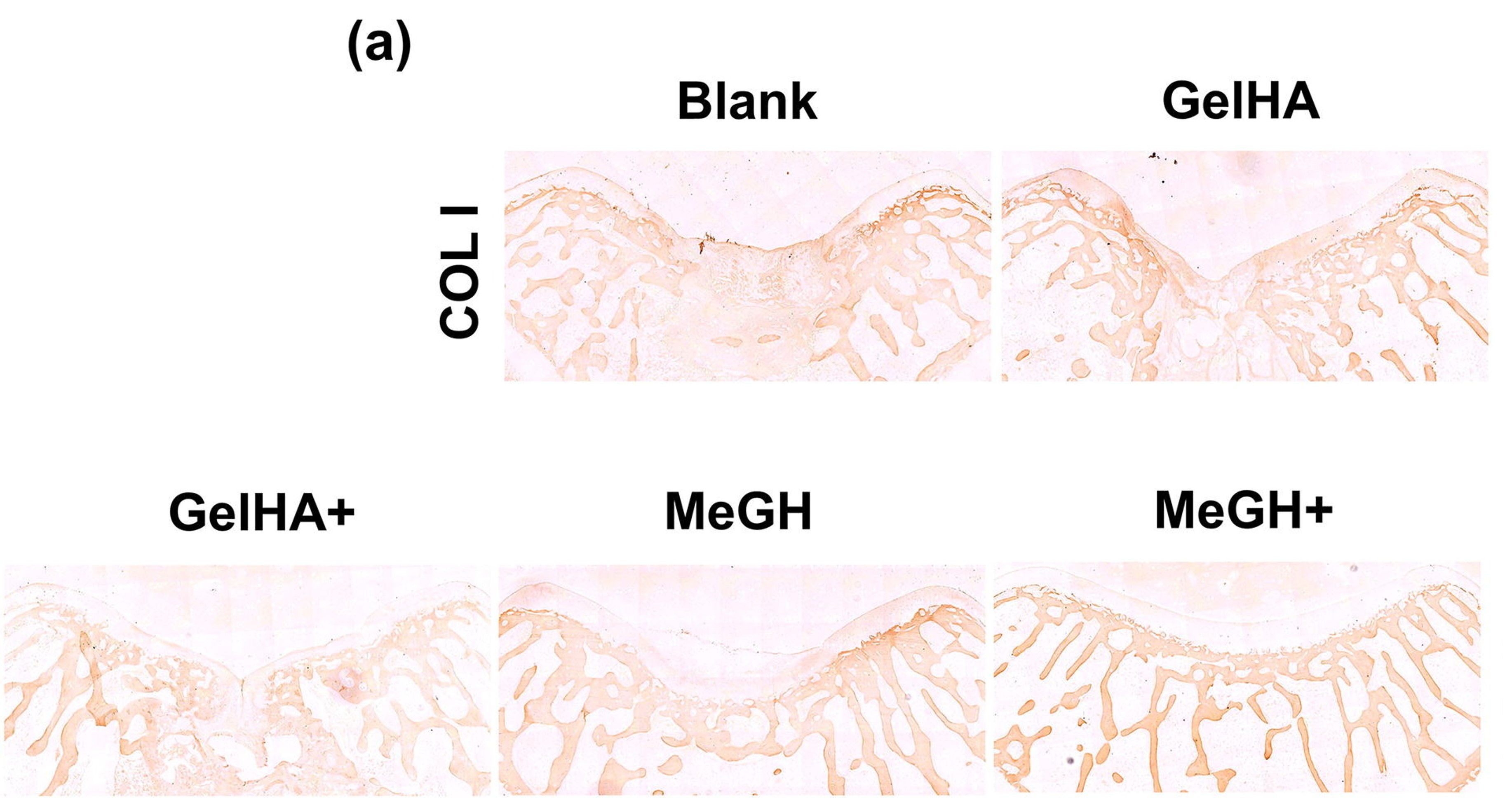
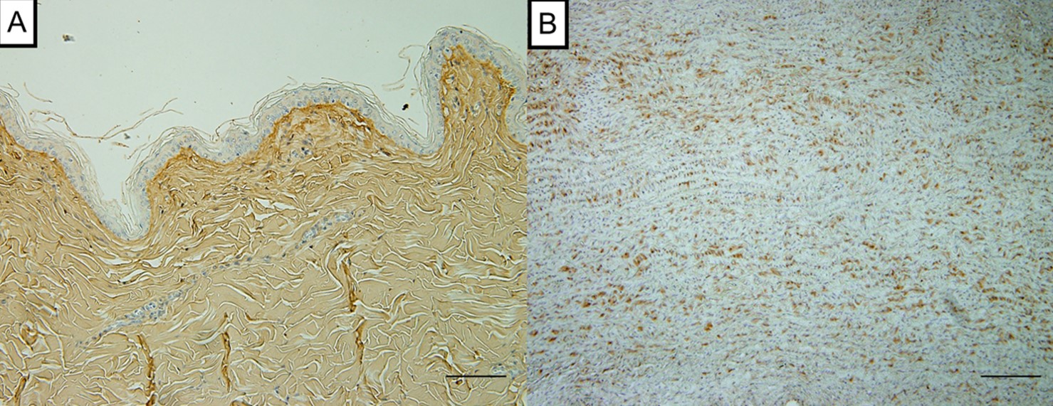
ARG21965 anti-Collagen I antibody, pre-adsorbed IHC-P image
Immunohistochemistry: Rabbit osteochondral stained with ARG21965 anti-Collagen I antibody, pre-adsorbed at 1:100 dilution.
From Wenli Dai et al. Enhanced osteochondral repair with hyaline cartilage formation using an extracellular matrix-inspired natural scaffold (2023), doi: 10.1016/j.scib.2023.07.050, Fig. 7a.
-
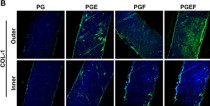
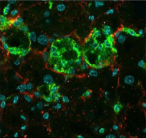
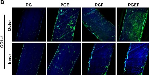
ARG21965 anti-Collagen I antibody, pre-adsorbed ICC/IF image
Immunofluorescence: Rabbit synovium-derived mesenchymal stem cell stained with ARG21965 anti-Collagen I antibody, pre-adsorbed at 1:1000 dilution.
From Zong Li e et al. Chemical Engineering Journal, (2023), doi: 10.1016/j.cej.2023.145209, Fig. 4B.
-
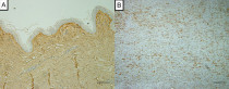
ARG21965 anti-Collagen I antibody, pre-adsorbed IHC-P image
Immunohistochemistry: Canine aorta mesothelial stained with ARG21965 anti-Collagen I antibody, pre-adsorbed at 1:500 dilution.
From Masakazu Shimada et al. PLoS One. (2022), doi: 35061786, Fig. 1.
-
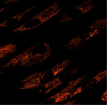
ARG21965 anti-Collagen I antibody, pre-adsorbed ICC/IF image
Immunofluorescence: Human fibroblasts were stained with ARG21965 anti-Collagen I antibody (pre-adsorbed).
-
ARG21965 anti-Collagen I antibody, pre-adsorbed IHC-Fr image
Immunohistochemistry: Frozen MC4R-KO Mouse liver section was stained with ARG21965 anti-Collagen I antibody (pre-adsorbed), anti-F4/80 antibody followed by secondary antibodies and DAPI.
-
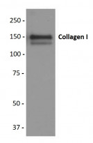
ARG21965 anti-Collagen I antibody, pre-adsorbed WB image
Western blot: Purified Human Type I Collagen stained with ARG21965 anti-Collagen I antibody (pre-adsorbed).
-
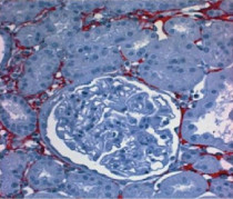
ARG21965 anti-Collagen I antibody, pre-adsorbed IHC-P image
Immunohistochemistry: Paraffin embedded Rat kidney section post uninephrectomy was stained with ARG21965 anti-Collagen I antibody (pre-adsorbed) followed by a secondary antibody and AEC.


Specific References
Tadalafil Ameliorates Chronic Ischemia-Associated Bladder Overactivity in Fructose-Fed Rats by Exerting Pelvic Angiogenesis and Enhancing p-eNOS Expression
WB / Rat


The protective effects of liraglutide in reducing lipid droplets accumulation and myocardial fibrosis in diabetic cardiomyopathy
WB / Rat
Effect of macrophage-to-myofibroblast transition on silicosis
ICC/IF, IHC-P,WB / Rat
Mesenchymal Stem Cells Preconditioned with Hypoxia and Dexamethasone Promote Osteoblast Differentiation Under Stress Conditions
ICC/IF / Human
Resveratrol attenuates inflammation and fibrosis in rheumatoid arthritis-associated interstitial lung disease via the AKT/TMEM175 pathway
WB, IHC-P / Human, Mouse
Bone marrow stromal and anterior cruciate ligament remnant cell co-culture-derived extracellular vesicles promote cell activity in both cell types
ICC/IF / Rabbit
Functional meniscus reconstruction with biological and biomechanical heterogeneities through topological self-induction of stem cells
ICC/IF / Rabbit
The potential of brimonidine for myopia treatment: Targeting MMP-2 to regulate choroidal thickness and control eye growth
IHC-Fr, WB / Guinea pig
Ultrasound-Controlled Delivery of Growth Factor-Loaded Cerasomes Combined with Polycaprolactone Scaffolds Seeded with Bone Marrow Mesenchymal Stem Cells for Biomimetic Tendon-to-Bone Interface Engineering
IHC-P / Rabbit
The mechano-chemical circuit in fibroblasts and dendritic cells drives basal cell proliferation in psoriasis
WB / Mouse
Effect of lyophilized exosomes derived from umbilical cord stem cells on chronic anterior cruciate ligament cell injury
ICC/IF / Human
Oxamate alleviates silicotic fibrosis in mice by inhibiting senescence of alveolar type II epithelial cells
WB / Mouse
Anti-Apoptosis Therapy for Meniscal Avascular Zone Repair: A Proof-of-Concept Study in a Lapine Model
IHC-P / Rabbit
Enhanced osteochondral repair with hyaline cartilage formation using an extracellular matrix-inspired natural scaffold
IHC-P / Rabbit
Regional specific tunable meniscus decellularized extracellular matrix (MdECM) reinforced bioink promotes anistropic meniscus regeneration
ICC/IF / Rabbit
Anterior cruciate ligament remnant preservation attenuates apoptosis and enhances the regeneration of hamstring tendon graft
ICC/IF / Rabbit
A Transwell-Based Vascularized Model to Investigate the Effect of Interstitial Flow on Vasculogenesis
ICC/IF / Human
Vinpocetine Ameliorates Metabolic-Syndrome-Associated Bladder Overactivity in Fructose-Fed Rats by Restoring Succinate-Modulated cAMP Levels and Exerting Anti-Inflammatory Effects in the Bladder Detrusor Muscle
WB / Mouse
The transplantation of particulated juvenile allograft cartilage and synovium for the repair of meniscal defect in a lapine model
IHC-P / Rabbit
Effects of long-term and high-dose administration of glucocorticoids on the cranial cruciate ligament in healthy beagle dogs
IHC-P / Dog
Loss of MLKL ameliorates liver fibrosis by inhibiting hepatocyte necroptosis and hepatic stellate cell activation
IHC-P / Mouse
Resveratrol Ameliorates Fibrosis in Rheumatoid Arthritis-Associated Interstitial Lung Disease via the Autophagy–Lysosome Pathway
WB / Mouse
Biphasic CK2.1-coated β-glycerophosphate chitosan/LL37-modified layered double hydroxide chitosan composite scaffolds enhance coordinated hyaline cartilage and subchondral bone regeneration.
IHC-P / Rabbit
hDPSC-laden GelMA microspheres fabricated using electrostatic microdroplet method for endodontic regeneration.
ICC/IF / Human
Effect of Freshly Isolated Bone Marrow Mononuclear Cells and Cultured Bone Marrow Stromal Cells in Graft Cell Repopulation and Tendon-Bone Healing after Allograft Anterior Cruciate Ligament Reconstruction.
IHC-P / Rabbit
Ba-Wei-Die-Huang-Wan (Hachimi-jio-gan) can ameliorate ketamine-induced cystitis by modulating neuroreceptors, inflammatory mediators, and fibrogenesis in a rat model.
WB / Rat
Activation of mTORC1 in fibroblasts accelerates wound healing and induces fibrosis in mice.
WB / Mouse
Stem cells from human exfoliated deciduous teeth as an alternative cell source in bio-root regeneration.
WB / Human


