ARG66162
anti-Fibronectin antibody
anti-Fibronectin antibody for IHC-Formalin-fixed paraffin-embedded sections,Western blot and Human,Mouse,Rat

Overview
| Product Description | Mouse Monoclonal antibody recognizes Fibronectin |
|---|---|
| Tested Reactivity | Hu, Ms, Rat |
| Tested Application | IHC-P, WB |
| Host | Mouse |
| Clonality | Monoclonal |
| Isotype | IgG |
| Target Name | Fibronectin |
| Antigen Species | Human |
| Immunogen | Synthetic peptide from Human Fibronectin |
| Conjugation | Un-conjugated |
| Alternate Names | ED-B; CIG; GFND; Cold-insoluble globulin; FNZ; LETS; GFND2; Fibronectin; MSF; FINC; FN |
Application Instructions
| Application Suggestion |
|
||||||
|---|---|---|---|---|---|---|---|
| Application Note | IHC-P: Antigen Retrieval: Boil tissue section in Sodium citrate buffer (pH 6.0) for 20 min. * The dilutions indicate recommended starting dilutions and the optimal dilutions or concentrations should be determined by the scientist. |
Properties
| Form | Liquid |
|---|---|
| Purification | Affinity purification with immunogen. |
| Buffer | PBS (pH 7.4), 0.02% Sodium azide and 50% Glycerol. |
| Preservative | 0.02% Sodium azide |
| Stabilizer | 50% Glycerol |
| Concentration | 1 mg/ml |
| Storage Instruction | For continuous use, store undiluted antibody at 2-8°C for up to a week. For long-term storage, aliquot and store at -20°C or below. Storage in frost free freezers is not recommended. Avoid repeated freeze/thaw cycles. Suggest spin the vial prior to opening. The antibody solution should be gently mixed before use. |
| Note | For laboratory research only, not for drug, diagnostic or other use. |
Bioinformation
| Database Links | |
|---|---|
| Gene Symbol | FN1 |
| Gene Full Name | fibronectin 1 |
| Background | This gene encodes fibronectin, a glycoprotein present in a soluble dimeric form in plasma, and in a dimeric or multimeric form at the cell surface and in extracellular matrix. Fibronectin is involved in cell adhesion and migration processes including embryogenesis, wound healing, blood coagulation, host defense, and metastasis. The gene has three regions subject to alternative splicing, with the potential to produce 20 different transcript variants. However, the full-length nature of some variants has not been determined. [provided by RefSeq, Jul 2008] |
| Function | Fibronectins bind cell surfaces and various compounds including collagen, fibrin, heparin, DNA, and actin. Fibronectins are involved in cell adhesion, cell motility, opsonization, wound healing, and maintenance of cell shape. Involved in osteoblast compaction through the fibronectin fibrillogenesis cell-mediated matrix assembly process, essential for osteoblast mineralization. Participates in the regulation of type I collagen deposition by osteoblasts. Anastellin binds fibronectin and induces fibril formation. This fibronectin polymer, named superfibronectin, exhibits enhanced adhesive properties. Both anastellin and superfibronectin inhibit tumor growth, angiogenesis and metastasis. Anastellin activates p38 MAPK and inhibits lysophospholipid signaling. [UniProt] |
| Highlight | Related Antibody Duos and Panels: ARG30346 Myofibroblast / Fibrosis Antibody Panel Related products: Fibronectin antibodies; Fibronectin ELISA Kits; Fibronectin Duos / Panels; Anti-Mouse IgG secondary antibodies; Related news: New antibody panels for Myofibroblasts and CAFs |
| Calculated MW | 272 kDa |
| PTM | Sulfated. It is not known whether both or only one of Thr-2064 and Thr-2065 are/is glycosylated. Forms covalent cross-links mediated by a transglutaminase, such as F13A or TGM2, between a glutamine and the epsilon-amino group of a lysine residue, forming homopolymers and heteropolymers (e.g. fibrinogen-fibronectin, collagen-fibronectin heteropolymers). Phosphorylated by FAM20C in the extracellular medium. Proteolytic processing produces the C-terminal NC1 peptide, anastellin. Some lysine residues are oxidized to allysine by LOXL3, promoting fibronectin activation and matrix formation. |
Images (9) Click the Picture to Zoom In
-
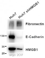
ARG66162 anti-Fibronectin antibody WB image
Western blot: 20 µg of Huh7 and Huh7 shHMGB1 cell lysates stained with ARG66162 anti-Fibronectin antibody (1:1000), ARG55914 anti-E-cadherin antibody (1:1000) and ARG65636 anti-HMGB1 antibody (1:2000).
-
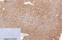
ARG66162 anti-Fibronectin antibody IHC-P image
Immunohistochemistry: Paraffin-embedded Rat liver tissue stained with ARG66162 anti-Fibronectin antibody at 1:200 (4°C, overnight). Antigen Retrieval: Boil tissue section in Sodium citrate buffer (pH 6.0) for 20 min. Secondary antibody was diluted at 1:200 (RT, 30min). Negative control: Secondary antibody only.
-
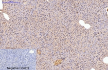
ARG66162 anti-Fibronectin antibody IHC-P image
Immunohistochemistry: Paraffin-embedded Mouse liver tissue stained with ARG66162 anti-Fibronectin antibody at 1:200 (4°C, overnight). Antigen Retrieval: Boil tissue section in Sodium citrate buffer (pH 6.0) for 20 min. Secondary antibody was diluted at 1:200 (RT, 30min). Negative control: Secondary antibody only.
-
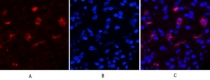
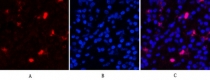
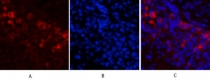
ARG66162 anti-Fibronectin antibody IHC image
Immunohistochemistry: Human appendix tissue stained with ARG66162 anti-Fibronectin antibody (red) at 1:200 (4°C, overnight). Picture A: Target. Picture B: DAPI. Picture C: merge of A and B.
-
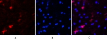
ARG66162 anti-Fibronectin antibody IHC image
Immunohistochemistry: Human appendix tissue stained with ARG66162 anti-Fibronectin antibody (red) at 1:200 (4°C, overnight). Picture A: Target. Picture B: DAPI. Picture C: merge of A and B.
-
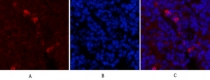
ARG66162 anti-Fibronectin antibody IHC image
Immunohistochemistry: Human appendix tissue stained with ARG66162 anti-Fibronectin antibody (red) at 1:200 (4°C, overnight). Picture A: Target. Picture B: DAPI. Picture C: merge of A and B.
-
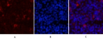
ARG66162 anti-Fibronectin antibody IHC image
Immunohistochemistry: Mouse spleen tissue stained with ARG66162 anti-Fibronectin antibody (red) at 1:200 (4°C, overnight). Picture A: Target. Picture B: DAPI. Picture C: merge of A and B.
-
ARG66162 anti-Fibronectin antibody IHC image
Immunohistochemistry: Mouse spleen tissue stained with ARG66162 anti-Fibronectin antibody (red) at 1:200 (4°C, overnight). Picture A: Target. Picture B: DAPI. Picture C: merge of A and B.
-
ARG66162 anti-Fibronectin antibody IHC image
Immunohistochemistry: Mouse spleen tissue stained with ARG66162 anti-Fibronectin antibody (red) at 1:200 (4°C, overnight). Picture A: Target. Picture B: DAPI. Picture C: merge of A and B.


Customer's Feedback
Good

Review for anti-Fibronectin antibody
Application:WB
Sample:Huh7 and Huh7 shHMGB1
Sample Loading Amount:20 µg
Primary Antibody Dilution Factor:1:1000
Primary Antibody Incubation Time:overnight
Primary Antibody Incubation Temperature:4 ºC

Specific References
Anticancer Effects and Molecular Mechanisms of Apigenin in Cervical Cancer Cells
WB, IHC-P / Human


