ARG30352
ISGF3 / GAF Antibody Panel
Component
| Cat No | Component Name | Host clonality | Reactivity | Application | Package |
|---|---|---|---|---|---|
| ARG43795 | anti-IRF9 antibody | Rabbit pAb | Hu | FACS, ICC/IF, WB | 20 μl |
| ARG51548 | anti-STAT1 phospho (Tyr701) antibody | Rabbit pAb | Hu, Ms, Rat | ICC/IF, IHC-P, WB | 20 μl |
| ARG66600 | anti-STAT2 phospho (Tyr690) antibody | Rabbit pAb | Hu, Ms, Rat | IHC-P, WB | 20 μg |
| ARG51041 | anti-STAT1 antibody | Rabbit pAb | Hu, Ms, Rat | IHC-P, WB | 20 μl |
| ARG57599 | anti-STAT2 antibody | Rabbit pAb | Hu, Ms, Rat | ICC/IF, IHC-P, WB | 20 μl |
Overview
| Product Description | Related news: ISGF3/GAF and JAK2-STAT3 antibody panels are launched |
|---|---|
| Target Name | ISGF3 / GAF Pathway Antibody |
Properties
| Storage Instruction | For continuous use, store undiluted antibody at 2-8°C for up to a week. For long-term storage, aliquot and store at -20°C or below. Storage in frost free freezers is not recommended. Avoid repeated freeze/thaw cycles. Suggest spin the vial prior to opening. The antibody solution should be gently mixed before use. |
|---|---|
| Note | For laboratory research only, not for drug, diagnostic or other use. |
Bioinformation
| Gene Full Name | Antibody Panel for ISGF3/GAF Pathway |
|---|---|
| Highlight | Related Product: anti-IRF9 antibody; anti-STAT1 phospho (Tyr701) antibody; anti-STAT2 phospho (Tyr690) antibody; anti-STAT1 antibody; anti-STAT2 antibody; |
Images (11) Click the Picture to Zoom In
-
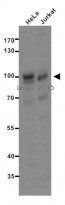
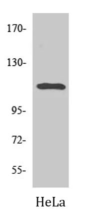
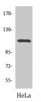
ARG51041 anti-STAT1 antibody WB image
Western blot: 20 µg of HeLa and Jurkat cell lysates stained with ARG51041 anti-STAT1 antibody at 1:500 dilution.
-
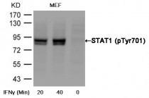
ARG51548 anti-STAT1 phospho (Tyr701) antibody WB image
Western blot: Extracts from MEF cells untreated or treated with interferon-ɤ (IFNɤ) stained with ARG51548 anti-STAT1 phospho (Tyr701) antibody.
-
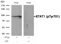
ARG51548 anti-STAT1 phospho (Tyr701) antibody WB image
Western blot: Extracts from HeLa cells, treated with IFNa or calf intestinal phosphatase (CIP), stained with ARG51548 anti-STAT1 phospho (Tyr701) antibody.
-
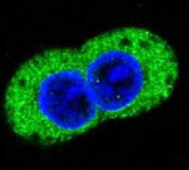
ARG57599 anti-STAT2 antibody ICC/IF image
Immunofluorescence: A431 cells stained with ARG57599 anti-STAT2 antibody.
-
ARG57599 anti-STAT2 antibody IHC-P image
Immunohistochemistry: Paraffin-embedded Human lung cancer tissue stained with ARG57599 anti-STAT2 antibody.
-

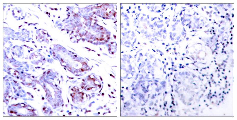
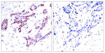
ARG66600 anti-STAT2 phospho (Tyr690) antibody IHC-P image
Immunohistochemistry: Paraffin-embedded Human lung carcinoma tissue stained with ARG66600 anti-STAT2 phospho (Tyr690) antibody. The picture on the right is blocked with the phosphopeptide.
-
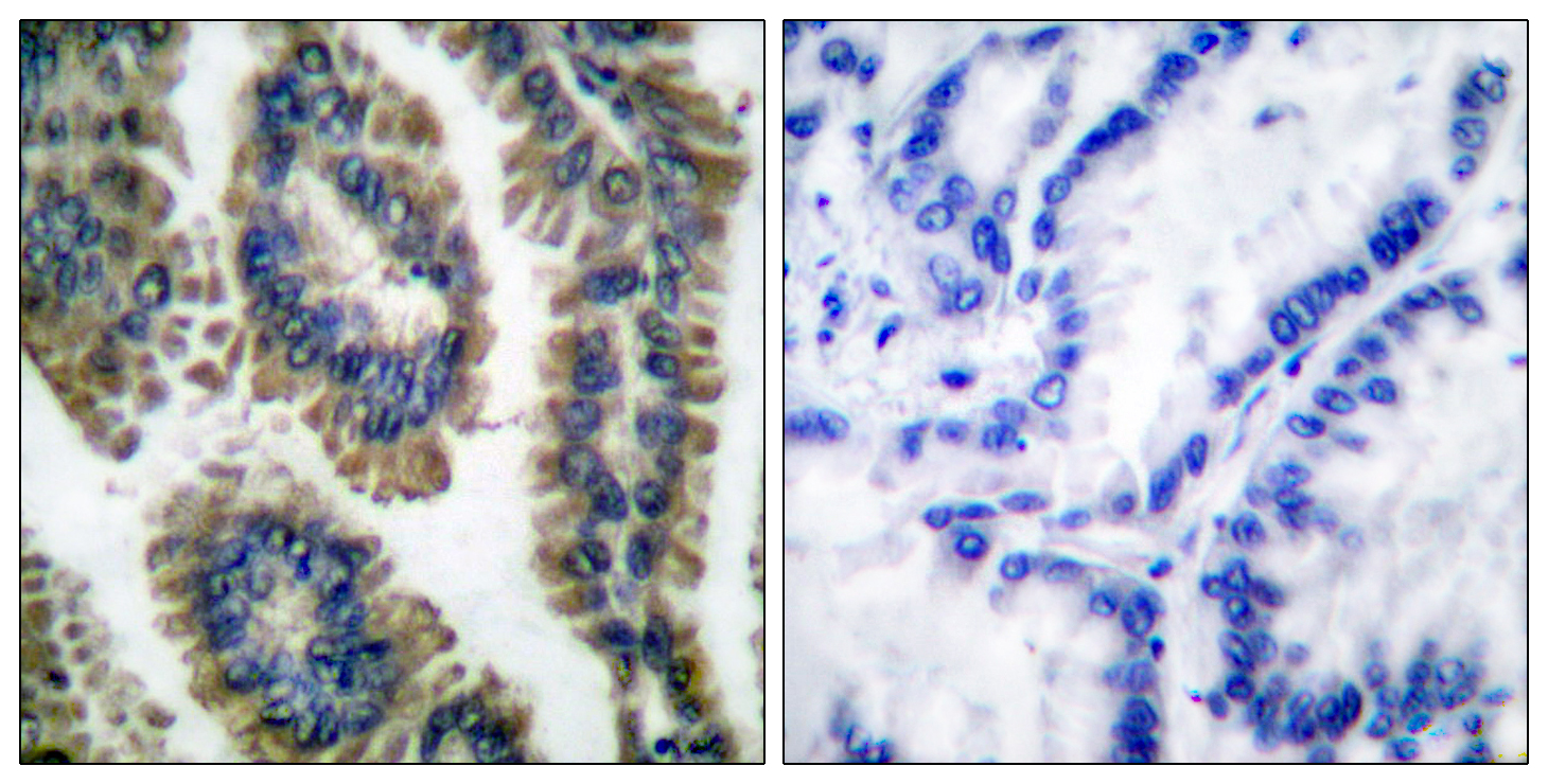
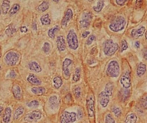
ARG51041 anti-STAT1 antibody IHC-P image
Immunohistochemistry: paraffin-embedded human breast carcinoma tissue stained with anti-STAT1 antibody ARG51041 (left) or the same antibody preincubated with blocking peptide (right).
-
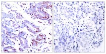
ARG51548 anti-STAT1 phospho (Tyr701) antibody IHC-P image
Immunohistochemistry: Paraffin-embedded Human breast carcinoma tissue stained with ARG51548 anti-STAT1 phospho (Tyr701) antibody (left) or the same antibody preincubated with blocking peptide (right).
-
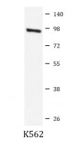
ARG57599 anti-STAT2 antibody WB image
Western blot: K562 cell lysate stained with ARG57599 anti-STAT2 antibody.
-
ARG66600 anti-STAT2 phospho (Tyr690) antibody WB image
Western blot: HeLa cell lysate stained with ARG66600 anti-STAT2 phospho (Tyr690) antibody.
-
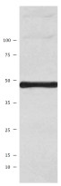
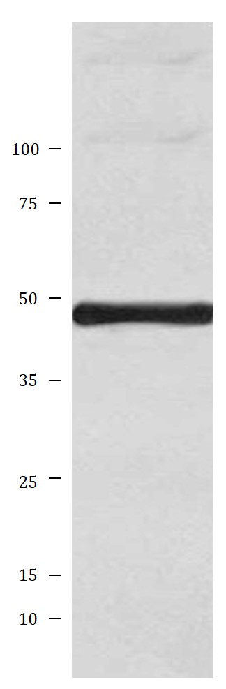
ARG43795 anti-IRF9 antibody WB image
Western blot: Jurkat cell lysate stained with ARG43795 anti-IRF9 antibody.