ARG41954
anti-CD279 / PD-1 antibody
anti-CD279 / PD-1 antibody for ICC/IF,Western blot and Human,Mouse,Rat
Overview
| Product Description | Rabbit Polyclonal antibody recognizes CD279 / PD-1 |
|---|---|
| Tested Reactivity | Hu, Ms, Rat |
| Tested Application | ICC/IF, WB |
| Host | Rabbit |
| Clonality | Polyclonal |
| Isotype | IgG |
| Target Name | CD279 / PD-1 |
| Antigen Species | Human |
| Immunogen | Synthetic peptide within aa. 1-100 of Human CD279 / PD-1 (NP_005009.2). |
| Conjugation | Un-conjugated |
| Alternate Names | hPD-l; CD279; PD-1; Protein PD-1; CD antigen CD279; PD1; hSLE1; SLEB2; Programmed cell death protein 1; hPD-1 |
Application Instructions
| Application Suggestion |
|
||||||
|---|---|---|---|---|---|---|---|
| Application Note | * The dilutions indicate recommended starting dilutions and the optimal dilutions or concentrations should be determined by the scientist. | ||||||
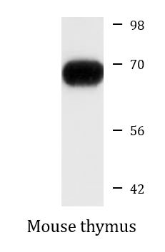
| Positive Control | Mouse thymus | ||||||
| Observed Size | ~ 68 kDa |
Properties
| Form | Liquid |
|---|---|
| Purification | Affinity purified. |
| Buffer | PBS (pH 7.3), 0.02% Sodium azide and 50% Glycerol. |
| Preservative | 0.02% Sodium azide |
| Stabilizer | 50% Glycerol |
| Storage Instruction | For continuous use, store undiluted antibody at 2-8°C for up to a week. For long-term storage, aliquot and store at -20°C. Storage in frost free freezers is not recommended. Avoid repeated freeze/thaw cycles. Suggest spin the vial prior to opening. The antibody solution should be gently mixed before use. |
| Note | For laboratory research only, not for drug, diagnostic or other use. |
Bioinformation
| Database Links | |
|---|---|
| Gene Symbol | PDCD1 |
| Gene Full Name | programmed cell death 1 |
| Background | CD279 / PD-1 is a cell surface membrane protein of the immunoglobulin superfamily. This protein is expressed in pro-B-cells and is thought to play a role in their differentiation. In mice, expression of this gene is induced in the thymus when anti-CD3 antibodies are injected and large numbers of thymocytes undergo apoptosis. Mice deficient for this gene bred on a BALB/c background developed dilated cardiomyopathy and died from congestive heart failure. These studies suggest that this gene product may also be important in T cell function and contribute to the prevention of autoimmune diseases. [provided by RefSeq, Jul 2008] |
| Function | CD279 / PD-1 is an inhibitory receptor on antigen activated T-cells. It plays a critical role in induction and maintenance of immune tolerance to self (PubMed:21276005). Delivers inhibitory signals upon binding to ligands CD274/PDCD1L1 and CD273/PDCD1LG2 (PubMed:21276005). Following T-cell receptor (TCR) engagement, PDCD1 associates with CD3-TCR in the immunological synapse and directly inhibits T-cell activation. Suppresses T-cell activation through the recruitment of PTPN11/SHP-2: following ligand-binding, PDCD1 is phosphorylated within the ITSM motif, leading to the recruitment of the protein tyrosine phosphatase PTPN11/SHP-2 that mediates dephosphorylation of key TCR proximal signaling molecules, such as ZAP70, PRKCQ/PKCtheta and CD247/CD3zeta. The PDCD1-mediated inhibitory pathway is exploited by tumors to attenuate anti-tumor immunity and escape destruction by the immune system, thereby facilitating tumor survival (PubMed:28951311). The interaction with CD274/PDCD1L1 inhibits cytotoxic T lymphocytes (CTLs) effector function (PubMed:28951311). The blockage of the PDCD1-mediated pathway results in the reversal of the exhausted T-cell phenotype and the normalization of the anti-tumor response, providing a rationale for cancer immunotherapy (PubMed:22658127, PubMed:25034862, PubMed:25399552). [UniProt] |
| Cellular Localization | Membrane; Single-pass type I membrane protein. [UniProt] |
| Highlight | Related products: PD-1 antibodies; PD-1 ELISA Kits; PD-1 Duos / Panels; Anti-Rabbit IgG secondary antibodies; Related news: The best solution for PD-1/PD-L1 research Examining CTL/NK-mediated cytotoxicity by ELISA |
| Calculated MW | 32 kDa |
Images (2) Click the Picture to Zoom In
-
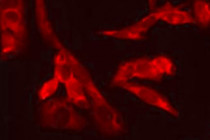
ARG41954 anti-CD279 / PD-1 antibody ICC/IF image
Immunofluorescence: U2OS cells stained with ARG41954 anti-CD279 / PD-1 antibody at 1:100 dilution.
-

ARG41954 anti-CD279 / PD-1 antibody WB image
Western blot: 25 µg of Mouse thymus lysate stained with ARG41954 anti-CD279 / PD-1 antibody at 1:1000 dilution.