ARG40204
anti-CD20 antibody
anti-CD20 antibody for Flow cytometry,IHC-Formalin-fixed paraffin-embedded sections,Western blot and Human
Cancer antibody; Developmental Biology antibody; Immune System antibody; B cell Marker antibody; Immature B Cell Marker antibody; Inflammatory Cell Marker antibody; Tumor-infiltrating Lymphocyte Study antibody
Overview
| Product Description | Rabbit Polyclonal antibody recognizes CD20 |
|---|---|
| Tested Reactivity | Hu |
| Tested Application | FACS, IHC-P, WB |
| Host | Rabbit |
| Clonality | Polyclonal |
| Isotype | IgG |
| Target Name | CD20 |
| Antigen Species | Human |
| Immunogen | Recombinant protein corresponding to M1-D261 of Human CD20. |
| Conjugation | Un-conjugated |
| Alternate Names | Bp35; LEU-16; B-lymphocyte surface antigen B1; B-lymphocyte antigen CD20; CD20; S7; CD antigen CD20; Leukocyte surface antigen Leu-16; B1; CVID5; Membrane-spanning 4-domains subfamily A member 1; MS4A2 |
Application Instructions
| Application Suggestion |
|
||||||||
|---|---|---|---|---|---|---|---|---|---|
| Application Note | IHC-P: Antigen Retrieval: Heat mediation was performed in Citrate buffer (pH 6.0) for 20 min. * The dilutions indicate recommended starting dilutions and the optimal dilutions or concentrations should be determined by the scientist. |
Properties
| Form | Liquid |
|---|---|
| Purification | Affinity purification with immunogen. |
| Buffer | 0.9% NaCl, 0.2% Na2HPO4, 0.05% Sodium azide and 5% BSA. |
| Preservative | 0.05% Sodium azide |
| Stabilizer | 5% BSA |
| Concentration | 0.5 mg/ml |
| Storage Instruction | For continuous use, store undiluted antibody at 2-8°C for up to a week. For long-term storage, aliquot and store at -20°C or below. Storage in frost free freezers is not recommended. Avoid repeated freeze/thaw cycles. Suggest spin the vial prior to opening. The antibody solution should be gently mixed before use. |
| Note | For laboratory research only, not for drug, diagnostic or other use. |
Bioinformation
| Database Links | |
|---|---|
| Gene Symbol | MS4A1 |
| Gene Full Name | membrane-spanning 4-domains, subfamily A, member 1 |
| Background | CD20 is a member of the membrane-spanning 4A gene family. Members of this nascent protein family are characterized by common structural features and similar intron/exon splice boundaries and display unique expression patterns among hematopoietic cells and nonlymphoid tissues. This gene encodes a B-lymphocyte surface molecule which plays a role in the development and differentiation of B-cells into plasma cells. This family member is localized to 11q12, among a cluster of family members. Alternative splicing of this gene results in two transcript variants which encode the same protein. [provided by RefSeq, Jul 2008] |
| Function | CD20 is a B-lymphocyte-specific membrane protein. It plays a role in the regulation of cellular calcium influx necessary for the development, differentiation, and activation of B-lymphocytes (PubMed:3925015, PubMed:7684739, PubMed:12920111). Functions as a store-operated calcium (SOC) channel component promoting calcium influx after activation by the B-cell receptor/BCR (PubMed:7684739, PubMed:12920111, PubMed:18474602). [UniProt] |
| Cellular Localization | Cell membrane; Multi-pass membrane protein. Cell membrane; Lipid-anchor. [UniProt] |
| Highlight | Related products: CD20 antibodies; CD20 ELISA Kits; CD20 Duos / Panels; Anti-Rabbit IgG secondary antibodies; Related news: New antibody panels and duos for Tumor immune microenvironment Tumor-Infiltrating Lymphocytes (TILs) Exploring Antiviral Immune Response |
| Research Area | Cancer antibody; Developmental Biology antibody; Immune System antibody; B cell Marker antibody; Immature B Cell Marker antibody; Inflammatory Cell Marker antibody; Tumor-infiltrating Lymphocyte Study antibody |
| Calculated MW | 33 kDa |
| PTM | Phosphorylated. Might be functionally regulated by protein kinase(s). [UniProt] |
Images (4) Click the Picture to Zoom In
-
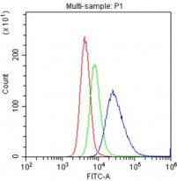
ARG40204 anti-CD20 antibody FACS image
Flow Cytometry: U937 cells were blocked with 10% normal goat serum and then stained with ARG40204 anti-CD20 antibody (blue) at 1 µg/10^6 cells for 30 min at 20°C, followed by DyLight®488 labelled secondary antibody. Isotype control antibody (green) was Rabbit IgG (1 µg/10^6 cells) used under the same conditions. Unlabelled sample (red) was also used as a control.
-
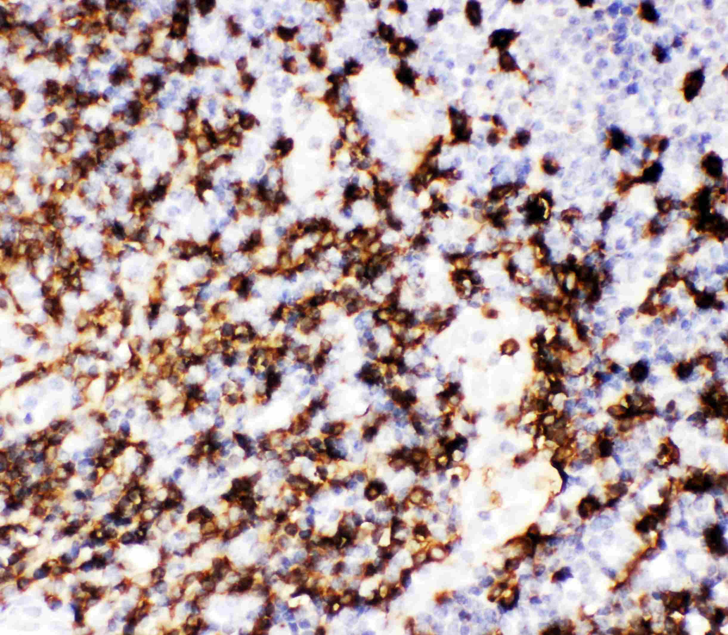
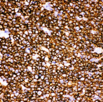
ARG40204 anti-CD20 antibody IHC-P image
Immunohistochemistry: Paraffin-embedded Human tonsil tissue. Antigen Retrieval: Heat mediation was performed in Citrate buffer (pH 6.0, epitope retrieval solution) for 20 min. The tissue section was blocked with 10% goat serum. The tissue section was then stained with ARG40204 anti-CD20 antibody at 1 µg/ml, overnight at 4°C.
-
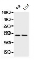
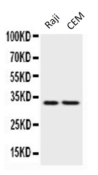
ARG40204 anti-CD20 antibody WB image
Western blot: 50 µg of samples under reducing conditions. Raji and CEM whole cell lysates stained with ARG40204 anti-CD20 antibody at 0.5 µg/ml, overnight at 4°C.
-
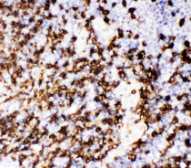
ARG40204 anti-CD20 antibody IHC-P image
Immunohistochemistry: Paraffin-embedded Human tonsil tissue. Antigen Retrieval: Heat mediation was performed in Citrate buffer (pH 6.0, epitope retrieval solution) for 20 min. The tissue section was blocked with 10% goat serum. The tissue section was then stained with ARG40204 anti-CD20 antibody at 1 µg/ml, overnight at 4°C.