ARG30334
Tumor-infiltrating Lymphocyte Antibody Panel
Tumor infiltrating Lymphocyte markers; Immunology; anti-tumor immune responses; tumor immune infiltration
Component
| Cat No | Component Name | Host clonality | Reactivity | Application | Package |
|---|---|---|---|---|---|
| ARG65859 | anti-CD3 epsilon antibody [SQab1713] | Rabbit mAb | Hu | FACS, ICC/IF, IHC-P, IP, WB | 20 μl |
| ARG66628 | anti-CD8 antibody [SQab19146] | Rabbit mAb | Hu | IHC-P | 20 μl |
| ARG65860 | anti-CD4 antibody [SQab1714] | Rabbit mAb | Hu | FACS, IHC-P, IP, WB | 20 μl |
| ARG66197 | anti-CD20 antibody [SQab1719] | Rabbit mAb | Hu | FACS, ICC/IF, IHC-P, IP | 20 μl |
Overview
| Product Description | Tumor-infiltrating Lymphocyte (TIL) Antibody Panel is an all-in-one solution to make identification of TILs easy and economic. This antibody panel comprises antibodies against CD3, CD4, CD8 and CD20 for identifying general T cells, Treg cells, cytotoxic T cells and B cells, respectively. All the antibodies in this panel have excellent IHC staining performance. Related news: New antibody panels and duos for Tumor immune microenvironment Tumor-Infiltrating Lymphocytes (TILs) |
|---|---|
| Target Name | Tumor-infiltrating Lymphocyte |
| Alternate Names | Tumor-infiltrating Lymphocyte antibody; CD3 antibody; CD4 antibody; CD20 antibody; CD8 antibody |
Properties
| Storage Instruction | For continuous use, store undiluted antibody at 2-8°C for up to a week. For long-term storage, aliquot and store at -20°C or below. Storage in frost free freezers is not recommended. Avoid repeated freeze/thaw cycles. Suggest spin the vial prior to opening. The antibody solution should be gently mixed before use. |
|---|---|
| Note | For laboratory research only, not for drug, diagnostic or other use. |
Bioinformation
| Gene Full Name | Antibody Panel for Tumor-infiltrating Lymphocyte |
|---|---|
| Highlight | Related Product: anti-CD3 epsilon antibody; anti-CD8 antibody; anti-CD4 antibody; anti-CD20 antibody; |
| Research Area | Tumor infiltrating Lymphocyte markers; Immunology; anti-tumor immune responses; tumor immune infiltration |
Images (17) Click the Picture to Zoom In
-
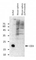
ARG65859 anti-CD3 epsilon antibody [SQab1713] WB image (Customer's Feedback)
Western blot: 20 µg of Jurkat and Mouse spleen (untreated or treated with LPS) lysates stained with ARG65859 anti-CD3 epsilon antibody [SQab1713] at 1:1000 dilution, overnight at 4°C.
-
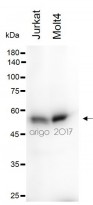
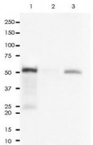
ARG65860 anti-CD4 antibody [SQab1714] WB image
Western blot: 30 µg of Jurkat and Molt4 cell lysates stained with ARG65860 anti-CD4 antibody [SQab1714] at 1:500 dilution.
-
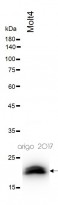
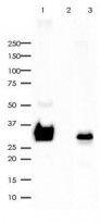
ARG65859 anti-CD3 epsilon antibody [SQab1713] WB image (Customer's Feedback)
Western blot: 30 µg of Molt4 cell lysate stained with ARG65859 anti-CD3 epsilon antibody [SQab1713] at 1:500 dilution.
-
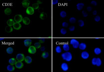
ARG65859 anti-CD3 epsilon antibody [SQab1713] ICC/IF image
Immunofluorescence: Jurkat cells were fixed with 4% paraformaldehyde for 30 min at RT, permeabilized with 0.1% Triton X-100 for 10 min at RT then blocked with 10% Goat serum for half an hour at room temperature. Samples were stained with ARG65859 anti-CD3 epsilon antibody [SQab1713] (green) at 1:50 and 4°C. DAPI (blue) was used as the nuclear counter stain. Control: PBS and secondary antibody.
-
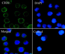
ARG66197 anti-CD20 antibody [SQab1719] ICC/IF image
Immunofluorescence: Raji cells fixed with 4% paraformaldehyde for 30 min at RT, permeabilized with 0.1% Triton X-100 for 10 min at RT then blocked with 10% Goat serum for half an hour at room temperature. Samples were stained with ARG66197 anti-CD20 antibody [SQab1719] (green) at 1:1000 at 4°C. DAPI (blue) was used as the nuclear counter stain. Control: PBS and secondary antibody.
-
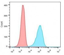
ARG65859 anti-CD3 epsilon antibody [SQab1713] FACS image
Flow Cytometry: Jurkat cells were fixed with 4% paraformaldehyde for 10 min. The cells were then stained with ARG65859 anti-CD3 epsilon antibody [SQab1713] (blue) at 1:1000 dilution in 1x PBS/1% BSA for 30 min at room temperture, followed by Alexa Fluor® 488 labelled secondary antibody. Unlabelled sample (red) was used as a control.
-
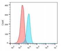
ARG65860 anti-CD4 antibody [SQab1714] FACS image
Flow Cytometry: Jurkat cells were fixed with 4% paraformaldehyde for 10 min. The cells were then stained with ARG65860 anti-CD4 antibody [SQab1714] (blue) at 1:50 dilution in 1x PBS/1% BSA for 30 min at room temperture, followed by Alexa Fluor® 488 labelled secondary antibody. Unlabelled sample (red) was used as a control.
-
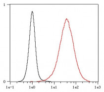
ARG66197 anti-CD20 antibody [SQab1719] FACS image
Flow Cytometry: Raji cells were fixed with 4% paraformaldehyde for 10 min. The cells were then stained with ARG66197 anti-CD20 antibody [SQab1719] (red) at 1:500 dilution in 1x PBS/1% BSA for 30 min at room temperture, followed by Alexa Fluor® 488 labelled secondary antibody. Unlabelled sample (black) was used as a control.
-
ARG65859 anti-CD3 epsilon antibody [SQab1713] IHC-P image
Immunohistochemistry: Formalin/PFA-fixed and paraffin-embedded sections of Human tonsil tissue stained with ARG65859 anti-CD3 epsilon antibody [SQab1713] at 1:200 dilution. Antigen Retrieval: Boil tissue section in Tris/EDTA buffer (pH 9.0).
-
ARG65860 anti-CD4 antibody [SQab1714] IHC-P image
Immunohistochemistry: Formalin/PFA-fixed and paraffin-embedded sections of Human tonsil tissue stained with ARG65860 anti-CD4 antibody [SQab1714] at 1:2000 dilution. Antigen Retrieval: Boil tissue section in Tris/EDTA buffer (pH 9.0).
-
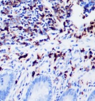
ARG66197 anti-CD20 antibody [SQab1719] IHC-P image
Immunohistochemistry: Formalin‐fixed and paraffin‐embedded Human appendix tissue stained with ARG66197 anti-CD20 antibody [SQab1719] at 1:20000 dilution.
Antigen retrieval: Heat mediated was performed using Tris/EDTA buffer pH 9.0
-

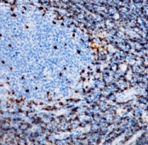
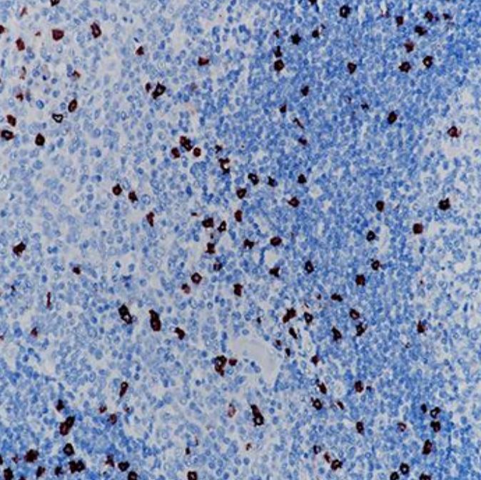
ARG66628 anti-CD8 antibody [SQab19146] IHC-P image
Immunohistochemistry: Formalin/PFA-fixed and paraffin-embedded Human tonsil tissue stained with ARG66628 anti-CD8 antibody [SQab19146]. Antigen Retrieval: Heat mediation was performed in Tris/EDTA buffer (pH 9.0).
-
ARG65859 anti-CD3 epsilon antibody [SQab1713] IP image
Immunoprecipitation: 0.4 mg of Molt-4 whole cell lysate was immunoprecipitated (1:15 dilution) and stained with ARG65859 anti-CD3 epsilon antibody [SQab1713].
Lane 1: Immunoprecipitation in Molt-4 whole cell lysate
Lane 2: Rabbit IgG instead of Primary Ab in Molt-4 whole cell lysate
Lane 3: Molt-4 whole cell lysate, 10 µg (input) -
ARG65860 anti-CD4 antibody [SQab1714] IP image
Immunoprecipitation: 0.4 mg of Molt-4 whole cell lysate was immunoprecipitated (1:50 dilution) and stained with ARG65860 anti-CD4 antibody [SQab1714].
Lane 1: Immunoprecipitation in Molt-4 whole cell lysate
Lane 2: Rabbit IgG instead of Primary Ab in Molt-4 whole cell lysate
Lane 3: Molt-4 whole cell lysate, 10 µg (input) -
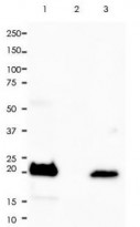
ARG66197 anti-CD20 antibody [SQab1719] IP image
Immunoprecipitation: 0.4 mg of Raji whole cell lysate was immunoprecipitated (1:20 dilution) and stained with ARG66197 anti-CD20 antibody [SQab1719].
Lane 1: Immunoprecipitation in Raji whole cell lysate
Lane 2: PBS instead of Primary Ab in Raji whole cell lysate
Lane 3: Raji whole cell lysate, 10 µg (input) -
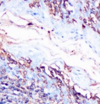
ARG65859 anti-CD3 epsilon antibody [SQab1713] IHC-P image
Immunohistochemistry: Formalin/PFA-fixed and paraffin-embedded sections of Human colon tissue stained with ARG65859 anti-CD3 epsilon antibody [SQab1713] at 1:200 dilution. Antigen Retrieval: Boil tissue section in Tris/EDTA buffer (pH 9.0).
-
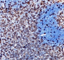
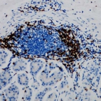
ARG66197 anti-CD20 antibody [SQab1719] IHC-P image
Immunohistochemistry: Formalin‐fixed and paraffin‐embedded Human spleen tissue stained with ARG66197 anti-CD20 antibody [SQab1719] at 1:20000 dilution.
Antigen retrieval: Heat mediated was performed using Tris/EDTA buffer pH 9.0


Specific References
Comparative transcriptomic analysis reveals differences in gene expression and regulatory pathways between nonacral and acral melanoma in Asian individuals
ARG66628: IHC-P / Human


