ARG30003
Tag Internal Control Antibody Duo (DDDK tag, beta Actin)
Controls and Markers antibody; Signaling Transduction antibody
Component
| Cat No | Component Name | Host clonality | Reactivity | Application | Package |
|---|---|---|---|---|---|
| ARG62986 | anti-DDDDK tag antibody [F-tag-01] | Mouse mAb | Other | ICC/IF, WB | 50 μg |
| ARG62346 | anti-beta Actin antibody [BA3R] | Mouse mAb | Hu, Ms, Rat, Chk, Rb | ELISA, FACS, ICC/IF, IP, WB | 50 μg |
Overview
| Product Description | DDDk tag, also known as FLAG-tag, FLAG octapeptide, DDDDK tag or DYKDDDDK tag, is a polypeptide protein tag (DYKDDDDK, 1012 Da) that can be added to a protein N or C-terminus using recombinant DNA technology. It can be used for affinity chromatography, to isolate recombinant protein from protein complexes with multiple subunits. DDDk tag is also used to detect protein cellular localization by immunofluorescence or protein expression level by SDS PAGE. Actins are a family of six proteins in three groups in human and other vertebrates: alpha ( ACTC1, cardiac muscle 1), alpha 1 ( ACTA1, skeletal muscle) and 2 ( ACTA2, aortic smooth muscle), beta ( ACTB), gamma 1 ( ACTG1) and 2 ( ACTG2, enteric smooth muscle). Beta and gamma 1 are two non-muscle actin proteins. They function as the main components of microfilaments. Actins are highly-conserved. Beta-actin proteins from species as diverse as human, mouse, and chicken are identical. Human beta actin shares close to 90% identity with fungal homologs. Human beta actin is at least 93% identical with other members of human actin family. Such homology information among the actin members and across species is important for antibody selection and the interpretation of Western blot bands. Both beta and alpha actins have been used as loading controls in Western blot experiments. [Labmoe] To study tagged-protein function in cells, researchers could use not only antibody recognizes tag polypeptide but also should chose a suitable internal control to control the loading of samples or the location of the intracellular organelles. ARG3003 Tag Internal control Duos (DDDK tag, beta Actin), provides not only an antibody reacts DDDK tag but also includes an antibody detects beta actin as a control to make your result more convincing |
|---|---|
| Target Name | Tag Internal Control |
| Alternate Names | Tag Internal Control antibody; beta Actin antibody; DDDDK tag antibody |
Properties
| Storage Instruction | For continuous use, store undiluted antibody at 2-8°C for up to a week. For long-term storage, aliquot and store at -20°C or below. Storage in frost free freezers is not recommended. Avoid repeated freeze/thaw cycles. Suggest spin the vial prior to opening. The antibody solution should be gently mixed before use. |
|---|---|
| Note | For laboratory research only, not for drug, diagnostic or other use. |
Bioinformation
| Gene Full Name | Antibody Duo for Tag Internal Control (DDDK tag, beta Actin) |
|---|---|
| Research Area | Controls and Markers antibody; Signaling Transduction antibody |
Images (7) Click the Picture to Zoom In
-
ARG62346 anti-beta Actin antibody [BA3R] ICC/IF image
Immunofluorescence: 100% Methanol fixed (RT, 10 min) HeLa cells stained with ARG62346 anti-beta Actin antibody [BA3R] at 1:500 dilution. Left: primary antibody (green). Right: Merge (primary antibody and DAPI).
Secondary antibody: ARG55393 Goat anti-Mouse IgG (H+L) antibody (FITC)
-
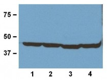
ARG62346 anti-beta Actin antibody [BA3R] WB image
Western Blot: 20 μg of (1) human, (2) mouse, (3) rat, and (4) rabbit tissue lysates stained with ARG62346 anti-Actin antibody [BA3R] at 1:1000 (1 μg/mL) dilution
-
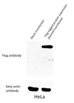
ARG62986 anti-DDDDK tag antibody [F-tag-01] WB image
Western blot: 1) Mock transfected 2) Flag-tagged protein expression plasmid transfected HeLa cell lysate stained with ARG62986 anti-DDDDK tag antibody [F-tag-01].
-
Monoclonal antibody clone F-tag-01 ICC/IF image
Immunofluorescence: COS-7 cells transfected with DDDDK expression vector stained with clone F-tag-01 followed by incubation with Goat anti-mouse IgG1 Alexa Fluor-594 (red). A) Fusion nuclear protein; B) cell nuclei stained with DAPI (blue); C) merged figures - confirmation of nuclear localization of the fusion protein; cell nuclei stained with DAPI (blue)
-
ARG62346 anti-beta Actin antibody [BA3R] WB image
Western blot: MCF-7, A549, H1299, HCT116, HepG2 and HUVEC cell lysates stained with ARG62346 anti-beta Actin antibody [BA3R] at 1:1000 dilution.
-
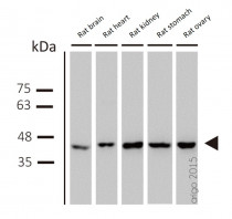
ARG62346 anti-beta Actin antibody [BA3R] WB image
Western blot: Rat brain, Rat heart, Rat kidney, Rat stomach and Rat ovary lysates stained with ARG62346 anti-beta Actin antibody [BA3R] at 1:1000 dilution.
-
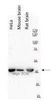
ARG62346 anti-beta Actin antibody [BA3R] WB image
Western blot: 20 µg of HeLa, Mouse brain and Rat brain lysates stained with ARG62346 anti-beta Actin antibody [BA3R] at 1:10000 dilution.
Specific References


