ARG30274
SOD1 and SOD2 Antibody Duo
Component
| Cat No | Component Name | Host clonality | Reactivity | Application | Package |
|---|---|---|---|---|---|
| ARG64241 | anti-SOD1 antibody | Goat pAb | Hu, Ms | ICC/IF, WB | 50 μg |
| ARG54937 | anti-SOD2 antibody | Rabbit pAb | Hu, Ms, Rat | ICC/IF, IHC-P, WB | 50 μl |
Overview
| Product Description | Superoxide Dismutases (SODs, including SOD1 and SOD2) are enzymes that alternately catalyzes the dismutation of superoxide radicals into less damaging molecular oxygen or hydrogen peroxide. The overproduction of superoxide radicals can cause severe tissue and cell damages. SOD enzymes deal with the superoxide radical by alternately adding or removing an electron from the superoxide molecules it encounters, thus changing the O2− into one of two less damaging species: either molecular oxygen (O2) or hydrogen peroxide (H2O2). SOD1 and SOD2 became the most important antioxidant defense systems in almost all living things exposed to oxygen. Fukui and Bao. 2010. Free Rad Biol and Med 6: 821-830 Nojima et al. 2015. Plos One 25(10) |
|---|---|
| Target Name | SOD1 and SOD2 |
| Alternate Names | SOD1 and SOD2 antibody; Superoxide dismutase 1 and Superoxide dismutase 2 antibody; SOD2 antibody; SOD1 antibody |
Properties
| Storage Instruction | For continuous use, store undiluted antibody at 2-8°C for up to a week. For long-term storage, aliquot and store at -20°C or below. Storage in frost free freezers is not recommended. Avoid repeated freeze/thaw cycles. Suggest spin the vial prior to opening. The antibody solution should be gently mixed before use. |
|---|---|
| Note | For laboratory research only, not for drug, diagnostic or other use. |
Bioinformation
| Gene Full Name | Superoxide dismutase 1 (SOD1) and Superoxide dismutase 2 (SOD2) Antibody Duo |
|---|---|
| Highlight | Related Product: anti-SOD1 antibody; anti-SOD2 antibody; |
Images (12) Click the Picture to Zoom In
-
ARG54937 anti-SOD2 antibody WB image
Western blot: Mouse hippocampal stained with ARG54937 anti-SOD2 antibody.
From Ji-Ren An et al. Front Cell Neurosci. (2023), doi: 10.3389/fncel.2023.1136070, Fig. 7F.
-
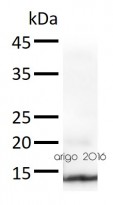
ARG64241 anti-SOD1 antibody WB image
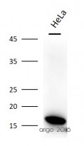
Western blot: 30 µg of HeLa cell lysate stained with ARG64241 anti-SOD1 antibody at 1:2000 dilution.
-
ARG54937 anti-SOD2 antibody ICC/IF image
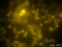
Immunofluorescence: 100% Methanol fixed (RT, 10 min) HeLa cells stained with ARG54937 anti-SOD2 antibody (orange) at 1:10 dilution.
Secondary antibody: ARG21917 Goat anti-Rabbit IgG antibody (TRITC)
-
ARG54937 anti-SOD2 antibody WB image
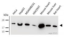
Western blot: 30 µg of HeLa, HepG2, MDAMB231, HEK293T, Mouse heart, Mouse liver, and Rat heart lysates stained with ARG54937 anti-SOD2 antibody at 1:500 dilution.
-
ARG64241 anti-SOD1 antibody WB image
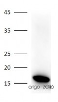
Western blot: 30 µg of HeLa cell lysate stained with ARG64241 anti-SOD1 antibody at 1:2000 dilution.
-
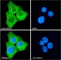
ARG64241 anti-SOD1 antibody ICC/IF image
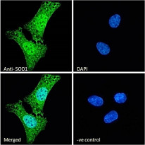
Immunofluorescence: Paraformaldehyde-fixed U2OS cells, permeabilized with 0.15% Triton. Cells were stained with ARG64241 anti-SOD1 antibody (green) at 10 µg/ml dilution for 1 hour. DAPI (blue) for nuclear staining. Negative control: Unimmunized Goat IgG (green) at 10 µg/ml dilution.
-
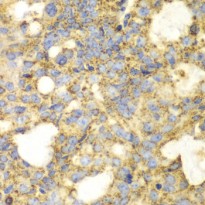
ARG54937 anti-SOD2 antibody IHC-P image
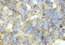
Immunohistochemistry: Paraffin-embedded Human oophoroma stained with ARG54937 anti-SOD2 antibody at 1:100 dilution (400x lens).
-
ARG54937 anti-SOD2 antibody IHC-P image
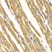
Immunohistochemistry: paraffin-embedded Rat heart stained with ARG54937 anti-SOD2 antibody at 1:100 dilution (400x lens).
-
ARG64241 anti-SOD1 antibody WB image
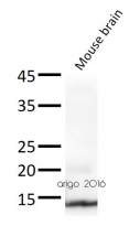
Western blot: 30 µg of Mouse brain lysate stained with ARG64241 anti-SOD1 antibody at 1:2000 dilution.
-
ARG64241 anti-SOD1 antibody WB image
Western blot: 30 µg of Mouse brain lysate stained with ARG64241 anti-SOD1 antibody at 1:2000 dilution.
-
ARG64241 anti-SOD1 antibody ICC/IF image
Immunofluorescence: Paraformaldehyde-fixed A431 cells, permeabilized with 0.15% Triton. Cells were stained with ARG64241 anti-SOD1 antibody (green) at 10 µg/ml dilution for 1 hour. DAPI (blue) for nuclear staining. Negative control: Unimmunized Goat IgG (green) at 10 µg/ml dilution.
-
ARG54937 anti-SOD2 antibody IHC-P image
Immunohistochemistry: paraffin-embedded Human oophoroma stained with ARG54937 anti-SOD2 antibody at 1:100 dilution (400x lens).
Specific References

