ARG64241
anti-SOD1 antibody
anti-SOD1 antibody for ICC/IF,Western blot and Human,Mouse
Cancer antibody; Cell Biology and Cellular Response antibody; Cell Death antibody; Gene Regulation antibody; Metabolism antibody; Microbiology and Infectious Disease antibody; Neuroscience antibody; Signaling Transduction antibody

Overview
| Product Description | Goat Polyclonal antibody recognizes SOD1 |
|---|---|
| Tested Reactivity | Hu, Ms |
| Predict Reactivity | Dog, Rat |
| Tested Application | ICC/IF, WB |
| Host | Goat |
| Clonality | Polyclonal |
| Isotype | IgG |
| Target Name | SOD1 |
| Antigen Species | Human |
| Immunogen | C-SRKHGGPKDEERH |
| Conjugation | Un-conjugated |
| Alternate Names | homodimer; EC 1.15.1.1; SOD; HEL-S-44; Superoxide dismutase [Cu-Zn]; ALS1; Superoxide dismutase 1; IPOA; ALS; hSod1 |
Application Instructions
| Application Suggestion |
|
||||||
|---|---|---|---|---|---|---|---|
| Application Note | WB: Recommend incubate at RT for 1h. * The dilutions indicate recommended starting dilutions and the optimal dilutions or concentrations should be determined by the scientist. |
Properties
| Form | Liquid |
|---|---|
| Purification | Purified from goat serum by antigen affinity chromatography. |
| Buffer | Tris saline (pH 7.3), 0.02% Sodium azide and 0.5% BSA. |
| Preservative | 0.02% Sodium azide |
| Stabilizer | 0.5% BSA |
| Concentration | 0.5 mg/ml |
| Storage Instruction | For continuous use, store undiluted antibody at 2-8°C for up to a week. For long-term storage, aliquot and store at -20°C or below. Storage in frost free freezers is not recommended. Avoid repeated freeze/thaw cycles. Suggest spin the vial prior to opening. The antibody solution should be gently mixed before use. |
| Note | For laboratory research only, not for drug, diagnostic or other use. |
Bioinformation
| Database Links | |
|---|---|
| Gene Symbol | SOD1 |
| Gene Full Name | superoxide dismutase 1, soluble |
| Background | The protein encoded by this gene binds copper and zinc ions and is one of two isozymes responsible for destroying free superoxide radicals in the body. The encoded isozyme is a soluble cytoplasmic protein, acting as a homodimer to convert naturally-occuring but harmful superoxide radicals to molecular oxygen and hydrogen peroxide. The other isozyme is a mitochondrial protein. Mutations in this gene have been implicated as causes of familial amyotrophic lateral sclerosis. Rare transcript variants have been reported for this gene. [provided by RefSeq, Jul 2008] |
| Highlight | Related Antibody Duos and Panels: ARG30274 SOD1 and SOD2 Antibody Duo Related products: SOD1 antibodies; SOD1 ELISA Kits; SOD1 Duos / Panels; Anti-Goat IgG secondary antibodies; Related poster download: The Structure & Functions of Mitochondria.pdf |
| Research Area | Cancer antibody; Cell Biology and Cellular Response antibody; Cell Death antibody; Gene Regulation antibody; Metabolism antibody; Microbiology and Infectious Disease antibody; Neuroscience antibody; Signaling Transduction antibody |
| Calculated MW | 16 kDa |
| PTM | Unlike wild-type protein, the pathogenic variants ALS1 Arg-38, Arg-47, Arg-86 and Ala-94 are polyubiquitinated by RNF19A leading to their proteasomal degradation. The pathogenic variants ALS1 Arg-86 and Ala-94 are ubiquitinated by MARCH5 leading to their proteasomal degradation. The ditryptophan cross-link at Trp-33 is responsible for the non-disulfide-linked homodimerization. Such modification might only occur in extreme conditions and additional experimental evidence is required. Palmitoylation helps nuclear targeting and decreases catalytic activity. Succinylation, adjacent to copper catalytic site, probably inhibits activity. Desuccinylation by SIRT5 enhances activity. |
Images (4) Click the Picture to Zoom In
-
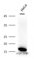
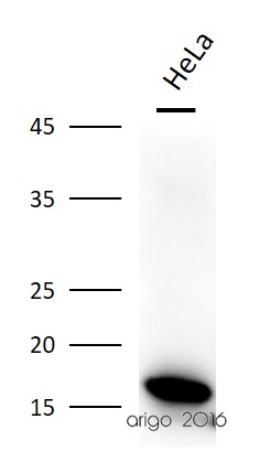
ARG64241 anti-SOD1 antibody WB image
Western blot: 30 µg of HeLa cell lysate stained with ARG64241 anti-SOD1 antibody at 1:2000 dilution.
-
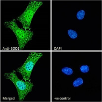
ARG64241 anti-SOD1 antibody ICC/IF image
Immunofluorescence: Paraformaldehyde-fixed U2OS cells, permeabilized with 0.15% Triton. Cells were stained with ARG64241 anti-SOD1 antibody (green) at 10 µg/ml dilution for 1 hour. DAPI (blue) for nuclear staining. Negative control: Unimmunized Goat IgG (green) at 10 µg/ml dilution.
-
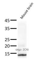
ARG64241 anti-SOD1 antibody WB image
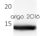
Western blot: 30 µg of Mouse brain lysate stained with ARG64241 anti-SOD1 antibody at 1:2000 dilution.
-
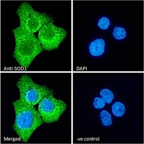
ARG64241 anti-SOD1 antibody ICC/IF image
Immunofluorescence: Paraformaldehyde-fixed A431 cells, permeabilized with 0.15% Triton. Cells were stained with ARG64241 anti-SOD1 antibody (green) at 10 µg/ml dilution for 1 hour. DAPI (blue) for nuclear staining. Negative control: Unimmunized Goat IgG (green) at 10 µg/ml dilution.
Customer's Feedback
