ARG30010
Neuronal Cytoskeletal Antibody Duo (NF-L, TUBBIII)
Controls and Markers antibody; Developmental Biology antibody; Neuroscience antibody; Signaling Transduction antibody
Component
| Cat No | Component Name | Host clonality | Reactivity | Application | Package |
|---|---|---|---|---|---|
| ARG52348 | anti-Neurofilament NF-L antibody [DA2] | Mouse mAb | Hu, Ms, Rat | FACS, ICC/IF, IHC-Fr, IHC-P, WB | 50 μl |
| ARG62683 | anti-beta III Tubulin antibody [TU-20] | Mouse mAb | Hu, Ms, Rat, Dog, Pig | FACS, ICC/IF, IHC-Fr, IHC-P, WB | 50 μg |
Overview
| Product Description | TuJ1/TUBBIII, a class III member of the beta tubulin protein family, is found in the cell bodies, dendrites, axons, and axonal terminations of immature neurons as a marker for immature neurons. NF-L, neurofilament light or low polypeptide, a 68-70 kDa protein, is a member of neurofilaments. Neurofilaments are a major component of the neuronal cytoskeleton, and are believed to function primarily to provide structural support for the axon and to regulate axon diameter. NF-L is as a marker for differentiated post-mitotic neuronal cells. ARG30010 Neuronal cytoskeletal Duos (NF-L, TUBBIII), including antibodies react two frequently used neuronal cytoskeletal target, NF-L and TUBBIII. This antibody duo not only provides neuronal cytoskeletal marker, but also inclueds neuron markers for differentiated neuronal and immature neuron which will be useful in neurogenesis, neural stem cell and neuronal disease studies. |
|---|---|
| Target Name | Neuronal Cytoskeletal |
| Alternate Names | Neuronal Cytoskeletal antibody; Neurofilament NF-L antibody; beta III Tubulin antibody |
Properties
| Storage Instruction | For continuous use, store undiluted antibody at 2-8°C for up to a week. For long-term storage, aliquot and store at -20°C or below. Storage in frost free freezers is not recommended. Avoid repeated freeze/thaw cycles. Suggest spin the vial prior to opening. The antibody solution should be gently mixed before use. |
|---|---|
| Note | For laboratory research only, not for drug, diagnostic or other use. |
Bioinformation
| Gene Full Name | Antibody Duo for Neuronal Cytoskeletal (NF-L, TUBBIII) |
|---|---|
| Highlight | Related Product: anti-Neurofilament NF-L antibody; anti-beta III Tubulin antibody; |
| Research Area | Controls and Markers antibody; Developmental Biology antibody; Neuroscience antibody; Signaling Transduction antibody |
Images (10) Click the Picture to Zoom In
-
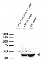
ARG62683 anti-beta III Tubulin antibody [TU-20] WB image
Western blot: 30 µg of 1) HeLa (negative control), 2) Mouse brain, and 3) Rat brain lysate stained with ARG62683 anti-beta III Tubulin antibody [TU-20] at 1:1000 dilution.
-
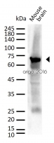
ARG52348 anti-Neurofilament NF-L antibody [DA2] WB image
Western blot: 30 µg of Mouse brain lysate stained with ARG52348 anti-Neurofilament NF-L antibody [DA2] at 1:1000 dilution.
-
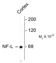
ARG52348 anti-Neurofilament NF-L antibody [DA2] WB image
Western blot: rat cortex lysate stained with ARG52348 anti-Neurofilament NF-L antibody [DA2] showing specific immunolableing of the ~ 68k NF-L protein.
-
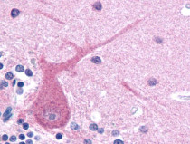
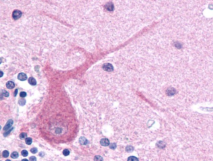
ARG62683 anti-beta III Tubulin antibody [TU-20] IHC-P image
Immunohistochemistry: Paraffin-embedded Human brain tissue stained with ARG62683 anti-beta III Tubulin antibody [TU-20].
-
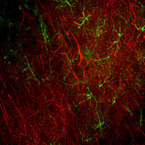
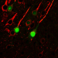
ARG52348 anti-Neurofilament NF-L antibody [DA2] IHC-Fr image
Immunohistochemistry: Frozen section of Rat frontal cortex tissue stained with ARG52348 anti-Neurofilament NF-L antibody [DA2] (red) at 1:500 dilution, and costained with anti-GFAP antibody (green) at 1:5000 dilution. Following transcardial perfusion of Rat with 4% paraformaldehyde, brain was post fixed for 24 hours, cut to 45 µM, and free-floating sections were stained with above antibodies.
Clone DA2 labels cell bodies and processes of pyramidal neurons, as well as dendrites and axons of other neuronal cells, while the GFAP antibody stains the network of glial cells.
-
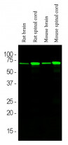
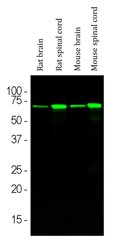
ARG52348 anti-Neurofilament NF-L antibody [DA2] WB image
Western blot: Rat brain, Rat spinal cord, Mouse brain and Mouse spinal cord lysates stained with ARG52348 anti-Neurofilament NF-L antibody [DA2] (green) at 1:5000 dilution.
-
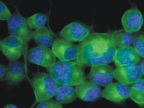
Monoclonal antibody clone TU-20 ICC/IF image
Immunofluorescence: Neuro2a mouse neuroblastoma cell stained with clone TU-20 (green)
Cell nuclei was stained with DAPI (blue). -
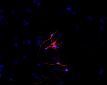
Monoclonal antibody clone TU-20 ICC/IF image
Immunofluorescence: P-19 mouse embryonal carcinoma cells stimulated to neuronal differentiation by retinoic acid stained with clone TU-20 (red)
Cell nuclei was stained with DAPI (blue). -
ARG62683 anti-beta III Tubulin antibody [TU-20] IHC-P image
Immunohistochemistry: Mouse brain stained with ARG62683 anti-beta III Tubulin antibody [TU-20].
-
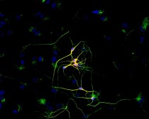
ARG62683 anti-beta III Tubulin antibody [TU-20] ICC/IF image
Immunofluorescence: P-19 mouse embryonal carcinoma cell line stimulated to neuronal differentiation by retinoic acid co-stained with stained with ARG62683 anti-beta III Tubulin antibody [TU-20] (red) and anti-beta-tubulin (green)
Superposition of red and green colours provided yellow staining. Nuclei were stained with DNA-binding dye (blue).


Specific References
Magnetic stirring with iron oxide nanospinners accretes neurotoxic Aβ42 oligomers into phagocytic clearable plaques for Alzheimer's disease treatment
ARG62683: ICC/IF / Mouse


