ARG30271
Mitochondrial Marker Antibody Panel (Cytochrome C, COX4, HSP60)
Component
| Cat No | Component Name | Host clonality | Reactivity | Application | Package |
|---|---|---|---|---|---|
| ARG54173 | anti-Hsp 60 antibody | Mouse mAb | Hu, Ms, Rat | ICC/IF, IP, WB | 50 μl |
| ARG54003 | anti-COX4 antibody | Mouse mAb | Hu, Ms, Rat, Goat, Hm, Mk | FACS, ICC/IF, IHC-P, IP, WB | 50 μl |
| ARG62461 | anti-Cytochrome C antibody [6H2.B4] | Mouse mAb | Hu, Ms, Rat | FACS, ICC/IF, IP, WB | 50 μl |
| ARG65350 | Goat anti-Mouse IgG antibody (HRP) | Goat pAb | Ms | ELISA, IHC-P, WB | 50 μl |
Overview
| Product Description | Cytochrome C, COX IV and HSP60 are markers for mitochondrial organelle. Cytochrome C is a component of the electron transport chain in mitochondria. The heme group of Cytochrome C accepts electrons from the b-c1 complex and transfers electrons to the cytochrome oxidase complex. HSP60 is a member of chaperon family. It is essential for the folding and assembly of newly imported proteins in the mitochondria. COX IV is one of the subunit of the Cytochrome C Oxidase (COX) hetero-oligomeric enzyme located in the inner mitochondrial membrane. It helps catalyzing the reduction of molecular oxygen to water coupled to the translocation of protons across inner membrane of mitochondria to drive ATP synthesis. Nishikimi et al. Nucleic Acids Res 16: 3577 (1988) Venner et al. DNA Cell Ciol 9: 545-552 (1990) Kadenbach et al. Free Radic Biol Med 29: 211-221 (2000) |
|---|---|
| Target Name | Mitochondrial Marker |
| Alternate Names | Mitochondrial Marker antibody; COX4 antibody; Hsp 60 antibody; Cytochrome C antibody |
Properties
| Storage Instruction | For continuous use, store undiluted antibody at 2-8°C for up to a week. For long-term storage, aliquot and store at -20°C or below. Storage in frost free freezers is not recommended. Avoid repeated freeze/thaw cycles. Suggest spin the vial prior to opening. The antibody solution should be gently mixed before use. |
|---|---|
| Note | For laboratory research only, not for drug, diagnostic or other use. |
Images (13) Click the Picture to Zoom In
-
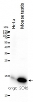
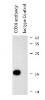
ARG62461 anti-Cytochrome C antibody [6H2.B4] WB image
Western blot: 20 µg of HeLa and Mouse testis lysates stained with ARG62461 anti-Cytochrome C antibody [6H2.B4] at 1:500 dilution.
-
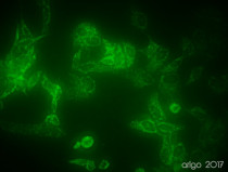
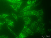
ARG54003 anti-COX4 antibody ICC/IF image
Immunofluorescence: 100% Methanol fixed (RT, 10 min) HeLa cells stained with ARG54003 anti-COX4 antibody (green) at 1:150 dilution.
Secondary antibody: ARG55393 Goat anti-Mouse IgG (H+L) antibody (FITC)
-
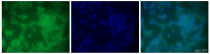
ARG54173 anti-Hsp 60 antibody ICC/IF image
Immunofluorescence: 100% Methanol fixed (RT, 10 min) HeLa cells stained with ARG54173 anti-Hsp 60 antibody at 1:100 dilution. Left: primary antibody (green). Middle: DAPI (blue). Right: Merge.
Secondary antibody: ARG55393 Goat anti-Mouse IgG (H+L) antibody (FITC)
-
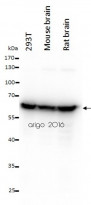
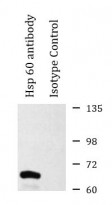
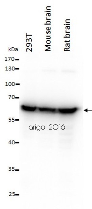
ARG54173 anti-Hsp 60 antibody WB image
Western blot: 20 µg of 293T, Mouse brain and Rat brain lysates stained with ARG54173 anti-Hsp 60 antibody at 1:1000 dilution.
-
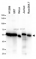
ARG54173 anti-Hsp 60 antibody WB image
Western blot: 15 µg of H1299, U87, HeLa, Jurkat and Raw264.7 cell lysates stained with ARG54173 anti-Hsp 60 antibody.
-
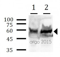
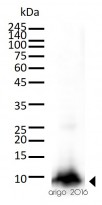
ARG54173 anti-Hsp60 antibody WB image
Western blot: 30 µg of 1) Mouse brain, and 2) Rat brain lysates stained with ARG54173 anti-Hsp60 antibody at 1:1000 dilution.
-
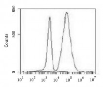
ARG54003 anti-COX4 antibody FACS image
Flow Cytometry: K562 cells stained with ARG54003 anti-COX4 antibody at 1:100 dilution (right histogram) or isotype control (left histogram), followed by incubation with FITC labelled secondary antibody.
-
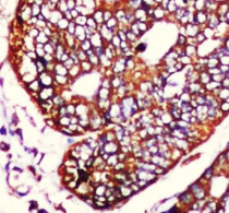
ARG54003 anti-COX4 antibody IHC-P image
Immunohistochemistry: Paraffin-embedded Human colorectal carcinoma stained with ARG54003 anti-COX4 antibody at 1:50 dilution. Antigen Retrieval: High-pressure and temperature Citrate buffer (pH 6.0).
-
ARG54003 anti-COX4 antibody IP image
Immunoprecipitation: HeLa cell lysates were immunoprecipitated and stained with ARG54003 anti-COX4 antibody.
-
ARG54173 anti-Hsp 60 antibody IP image
Immunoprecipitation: HeLa cell lysates were immunoprecipitated and stained with ARG54173 anti-Hsp 60 antibody.
-
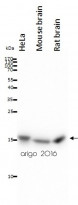
ARG54003 anti-COX4 antibody WB image
Western blot: 20 µg of HeLa, Mouse brain and Rat brain lysates stained with ARG54003 anti-COX4 antibody at 1:1000 dilution.
-
ARG62461 anti-Cytochrome C antibody [6H2.B4] WB image
Western blot: 30 µg of Mouse kidney lysate stained with ARG62461 anti-Cytochrome C antibody [6H2.B4] at 1:500 dilution.
-
ARG54003 anti-COX4 antibody ICC/IF image
Immunofluorescence: 100% Methanol fixed (RT, 10 min) HeLa cells stained with ARG54003 anti-COX4 antibody (green) at 1:150 dilution.
Secondary antibody: ARG55393 Goat anti-Mouse IgG (H+L) antibody (FITC)


Specific References
KDF1 Promoted Proliferation, Migration and Invasion of Lung Adenocarcinoma Cells through Activating STAT3 and AKT Pathway
ARG65350: WB /


RSV replication is promoted by autophagy-mediated inhibition of apoptosis.
ARG54173: WB / Human


