ARG54003
anti-COX4 antibody
anti-COX4 antibody for Flow cytometry,ICC/IF,IHC-Formalin-fixed paraffin-embedded sections,Immunoprecipitation,Western blot and Human,Mouse,Rat,Goat,Hamster,Monkey
Cancer antibody; Controls and Markers antibody; Metabolism antibody; Signaling Transduction antibody; Loading Control antibody for Cytoplasmic Fractions; Cytochrome-C fractionation Study antibody; Mitochondrial Marker antibody

Overview
| Product Description | Mouse Monoclonal antibody recognizes COX4 |
|---|---|
| Tested Reactivity | Hu, Ms, Rat, Goat, Hm, Mk |
| Tested Application | FACS, ICC/IF, IHC-P, IP, WB |
| Host | Mouse |
| Clonality | Monoclonal |
| Isotype | IgG1 |
| Target Name | COX4 |
| Antigen Species | Human |
| Immunogen | A synthetic peptide corresponding to carboxyl terminal residues of human COX IV |
| Conjugation | Un-conjugated |
| Alternate Names | Cytochrome c oxidase polypeptide IV; COX4; COX4-1; COXIV; Cytochrome c oxidase subunit IV isoform 1; COX IV-1; Cytochrome c oxidase subunit 4 isoform 1, mitochondrial |
Application Instructions
| Application Suggestion |
|
||||||||||||
|---|---|---|---|---|---|---|---|---|---|---|---|---|---|
| Application Note | IHC-P: Antigen Retrieval: High-pressure and temperature Citrate buffer (pH 6.0). * The dilutions indicate recommended starting dilutions and the optimal dilutions or concentrations should be determined by the scientist. |
Properties
| Form | Liquid |
|---|---|
| Purification | Affinity purified |
| Buffer | 0.1M Tris-Glycine (pH 7.4), 150 mM NaCl, 0.2% Sodium azide, 0.1mg/ml BSA and 50% Glycerol |
| Preservative | 0.2% Sodium azide |
| Stabilizer | 0.1mg/ml BSA, 50% Glycerol |
| Concentration | 1 mg/ml |
| Storage Instruction | For continuous use, store undiluted antibody at 2-8°C for up to a week. For long-term storage, aliquot and store at -20°C. Storage in frost free freezers is not recommended. Avoid repeated freeze/thaw cycles. Suggest spin the vial prior to opening. The antibody solution should be gently mixed before use. |
| Note | For laboratory research only, not for drug, diagnostic or other use. |
Bioinformation
| Database Links | |
|---|---|
| Gene Symbol | COX4I1 |
| Gene Full Name | cytochrome c oxidase subunit IV isoform 1 |
| Background | Cytochrome c oxidase (COX) is the terminal enzyme of the mitochondrial respiratory chain. It is a multi-subunit enzyme complex that couples the transfer of electrons from cytochrome c to molecular oxygen and contributes to a proton electrochemical gradient across the inner mitochondrial membrane. The complex consists of 13 mitochondrial- and nuclear-encoded subunits. The mitochondrially-encoded subunits perform the electron transfer and proton pumping activities. The functions of the nuclear-encoded subunits are unknown but they may play a role in the regulation and assembly of the complex. This gene encodes the nuclear-encoded subunit IV isoform 1 of the human mitochondrial respiratory chain enzyme. It is located at the 3' of the NOC4 (neighbor of COX4) gene in a head-to-head orientation, and shares a promoter with it. Pseudogenes related to this gene are located on chromosomes 13 and 14. Alternative splicing results in multiple transcript variants encoding different isoforms. [provided by RefSeq, Jan 2016] |
| Function | COX4 protein is one of the nuclear-coded polypeptide chains of cytochrome c oxidase, the terminal oxidase in mitochondrial electron transport. [UniProt] |
| Cellular Localization | Mitochondrion inner membrane. [UniProt] |
| Highlight | Related Antibody Duos and Panels: ARG30259 Loading Controls for Cytoplasmic / Nuclear Fractions Antibody Panel ARG30271 Mitochondrial Marker Antibody Panel (Cytochrome C, COX4, HSP60) ARG30276 Cytochrome-C fractionation Antibody Panel (Cytochrome-C, COX IV, beta Actin) Related products: COX IV antibodies; COX IV Duos / Panels; Anti-Mouse IgG secondary antibodies; Related poster download: The Structure & Functions of Mitochondria.pdf |
| Research Area | Cancer antibody; Controls and Markers antibody; Metabolism antibody; Signaling Transduction antibody; Loading Control antibody for Cytoplasmic Fractions; Cytochrome-C fractionation Study antibody; Mitochondrial Marker antibody |
| Calculated MW | 20 kDa |
Images (6) Click the Picture to Zoom In
-
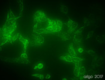
ARG54003 anti-COX4 antibody ICC/IF image
Immunofluorescence: 100% Methanol fixed (RT, 10 min) HeLa cells stained with ARG54003 anti-COX4 antibody (green) at 1:150 dilution.
Secondary antibody: ARG55393 Goat anti-Mouse IgG (H+L) antibody (FITC)
-
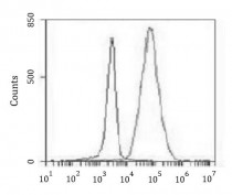
ARG54003 anti-COX4 antibody FACS image
Flow Cytometry: K562 cells stained with ARG54003 anti-COX4 antibody at 1:100 dilution (right histogram) or isotype control (left histogram), followed by incubation with FITC labelled secondary antibody.
-
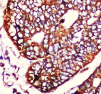
ARG54003 anti-COX4 antibody IHC-P image
Immunohistochemistry: Paraffin-embedded Human colorectal carcinoma stained with ARG54003 anti-COX4 antibody at 1:50 dilution. Antigen Retrieval: High-pressure and temperature Citrate buffer (pH 6.0).
-
ARG54003 anti-COX4 antibody IP image
Immunoprecipitation: HeLa cell lysates were immunoprecipitated and stained with ARG54003 anti-COX4 antibody.
-
ARG54003 anti-COX4 antibody WB image
Western blot: 20 µg of HeLa, Mouse brain and Rat brain lysates stained with ARG54003 anti-COX4 antibody at 1:1000 dilution.
-
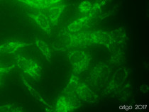
ARG54003 anti-COX4 antibody ICC/IF image
Immunofluorescence: 100% Methanol fixed (RT, 10 min) HeLa cells stained with ARG54003 anti-COX4 antibody (green) at 1:150 dilution.
Secondary antibody: ARG55393 Goat anti-Mouse IgG (H+L) antibody (FITC)
Customer's Feedback
 Excellent
Review for anti-COX4 antibody
Application:IF/ICC
Sample:HeLa
Fixation Buffer:100% Methanol
Fixation Time:10 min
Fixation Temperature:RT ºC
Permeabilization Buffer:0.1% Triton X-100
Primary Antibody Dilution Factor:1:150
Primary Antibody Incubation Time:overnight
Primary Antibody Incubation Temperature:4 ºC
Conjugation of Secondary Antibody:FITC


