ARG30258
Loading Controls for Whole Cell Lysate Antibody Panel
Cancer antibody; Cell Biology and Cellular Response antibody; Controls and Markers antibody; Immune System antibody; Metabolism antibody; Signaling Transduction antibody
Component
| Cat No | Component Name | Host clonality | Reactivity | Application | Package |
|---|---|---|---|---|---|
| ARG65714 | anti-Vinculin antibody | Rabbit pAb | Hu, Ms, Rat | WB | 25 μl |
| ARG65693 | anti-alpha Tubulin antibody | Rabbit pAb | Hu, Ms, Rat | FACS, ICC/IF, IHC-P, WB | 25 μl |
| ARG65683 | anti-beta Actin antibody | Rabbit pAb | Hu, Ms, Rat, Rb, Sheep | IHC-P, WB | 25 μg |
| ARG65680 | anti-GAPDH antibody | Rabbit pAb | Hu, Ms, Rat | ICC/IF, IHC-P, WB | 25 μl |
| ARG65351 | Goat anti-Rabbit IgG antibody (HRP) | Goat pAb | Rb | ELISA, IHC-P, WB | 50 μl |
Overview
| Target Name | Loading Controls for Whole Cell Lysate |
|---|---|
| Alternate Names | Loading Controls for Whole Cell Lysate antibody; GAPDH antibody; beta Actin antibody; alpha Tubulin antibody; Vinculin antibody |
Properties
| Storage Instruction | For continuous use, store undiluted antibody at 2-8°C for up to a week. For long-term storage, aliquot and store at -20°C or below. Storage in frost free freezers is not recommended. Avoid repeated freeze/thaw cycles. Suggest spin the vial prior to opening. The antibody solution should be gently mixed before use. |
|---|---|
| Note | For laboratory research only, not for drug, diagnostic or other use. |
Bioinformation
| Gene Full Name | Antibody Panel for Loading Controls for Whole Cell Lysate |
|---|---|
| Highlight | Related Product: anti-Vinculin antibody; anti-alpha Tubulin antibody; anti-beta Actin antibody; anti-GAPDH antibody; Organelle Marker; Loading Control; |
| Research Area | Cancer antibody; Cell Biology and Cellular Response antibody; Controls and Markers antibody; Immune System antibody; Metabolism antibody; Signaling Transduction antibody |
Images (35) Click the Picture to Zoom In
-
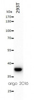
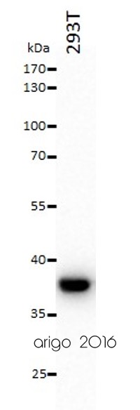
ARG65680 anti-GAPDH antibody WB image
Western blot: 20 µg of 293T cell lysate stained with ARG65680 anti-GAPDH antibody at 1:10000 dilution.
-
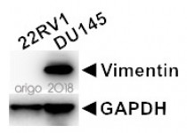
ARG65680 anti-GAPDH antibody WB image
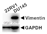
Western blot: 20 µg of 22RV1 and DU145 cell lysates stained with ARG66302 anti-Vimentin antibody [SQab1859] at 1:2000 dilution and ARG65680 anti-GAPDH antibody at 1:10000 dilution.
-

ARG65680 anti-GAPDH antibody WB image
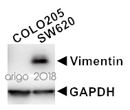
Western blot: 20 µg of COLO205 and SW620 cell lysates stained with ARG66302 anti-Vimentin antibody [SQab1859] at 1:2000 dilution and ARG65680 anti-GAPDH antibody at 1:10000 dilution.
-
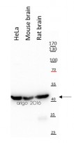
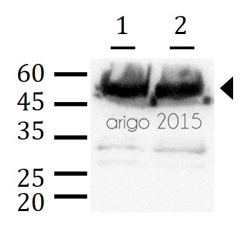
ARG65683 anti-beta Actin antibody WB image
Western blot: 20 µg of HeLa, Mouse brain and Rat brain lysates stained with ARG65683 anti-beta Actin antibody at 1:10000 dilution.
-
ARG65683 anti-beta Actin antibody WB image
Western blot: 30 µg of 293T lysate stained with ARG65683 anti-beta Actin antibody at 1:3000 dilution.
-
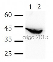
ARG65683 anti-beta Actin antibody WB image
Western blot: 30 µg of 1) Rat brain, and 2) Mouse liver lysate stained with ARG65683 anti-beta Actin antibody at 1:3000 dilution.
-
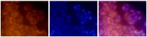
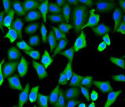
ARG65693 anti-alpha Tubulin antibody ICC/IF image
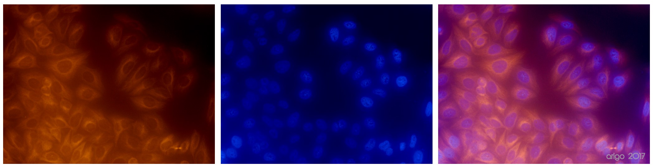
Immunofluorescence: 100% Methanol fixed (RT, 10 min) HeLa cells stained with ARG65693 anti-alpha Tubulin antibody at 1:200 dilution. Left: primary antibody (orange). Middle: DAPI (blue). Right: Merge.
Secondary antibody: ARG21917 Goat anti-Rabbit IgG antibody (TRITC)
-
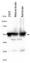
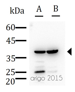
ARG65693 anti-alpha Tubulin antibody WB image
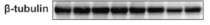
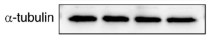
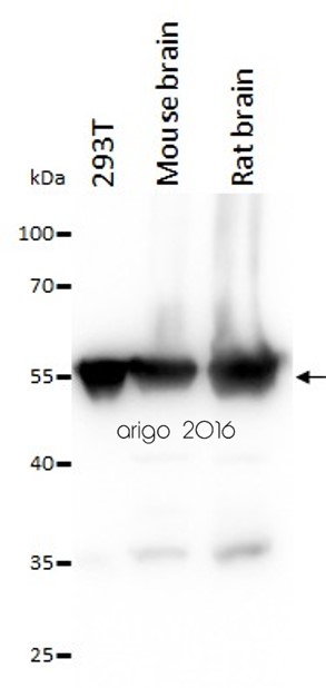
Western blot: 20 µg of 293T, Mouse brain and Rat brain lysates stained with ARG65693 anti-alpha Tubulin antibody at 1:10000 dilution.
-

ARG65693 anti-alpha Tubulin antibody WB image
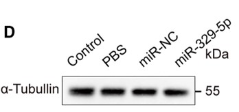
Western blot: Mouse retina stained with ARG65693 anti-alpha Tubulin antibody and ARG65351 Goat anti-Rabbit IgG antibody (HRP).
From Xiaoyuan Ye et al. Mol Ther Nucleic Acids. (2024), doi: 10.1016/j.omtn.2024.102209, Fig. 5.D.
-
-
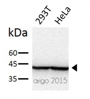
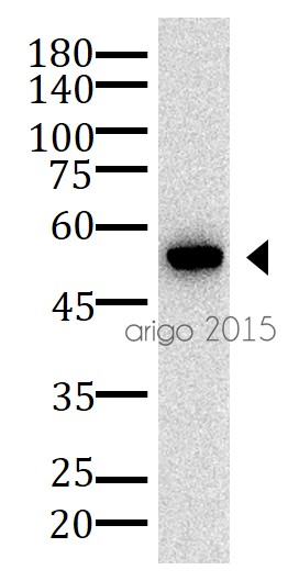
ARG65680 anti-GAPDH antibody WB image
Western blot: 30 µg of HeLa cell lysate stained with ARG65680 anti-GAPDH antibody at 1:500 dilution.
-

ARG65680 anti-GAPDH antibody ICC/IF image
Immunofluorescence: MCF-7 stained with ARG65680 anti-GAPDH antibody. Blue: DAPI for nuclear staining.
-

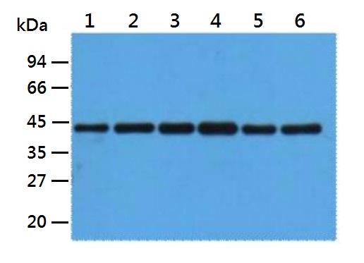
ARG65683 anti-beta Actin antibody WB image
Western blot: 1) HepG2, 2) Rat liver, 3) Mouse kidney, 4) Rabbit testis, 5) Sheep lung, and 6) 293T lysates stained with ARG65683 anti-beta Actin antibody at 1:5000 dilution.
-
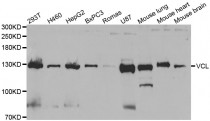
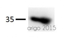
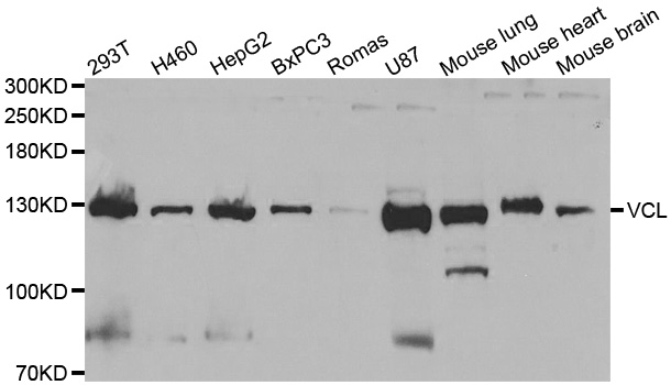
ARG65714 anti-Vinculin antibody WB image
Western blot: extracts of various cell lines stained with ARG65714 anti-Vinculin antibody.
-
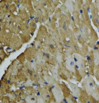
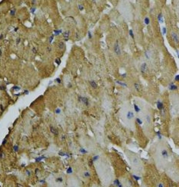
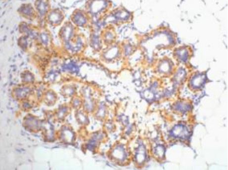
ARG65680 anti-GAPDH antibody IHC-P image
Immunohistochemistry: Paraffin-embedded Mouse heart stained with ARG65680 anti-GAPDH antibody at 1:100 dilution.
-
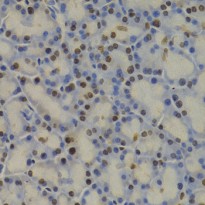
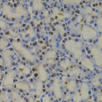
ARG65680 anti-GAPDH antibody IHC-P image
Immunohistochemistry: paraffin-embedded Rat pancreas stained with ARG65680 anti-GAPDH antibody at 1:100 dilution (400x lens).
-

ARG65683 anti-beta Actin antibody IHC-P image
Immunohistochemistry: Human ovary tissue stained with ARG65683 anti-beta Actin antibody at 1:200 dilution.
-
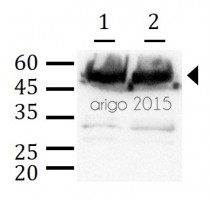
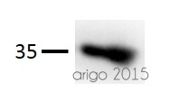
ARG65693 anti-alpha Tubulin antibody WB image
Western blot: 30 µg of HeLa cell lysate stained with ARG65693 anti-alpha Tubulin antibody at 1:5000 dilution.
-
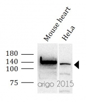
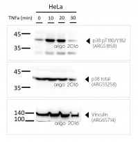
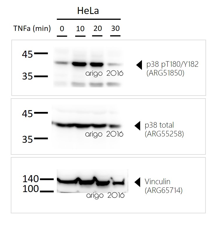
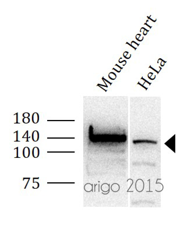
ARG65714 anti-Vinculin antibody WB image
Western blot: 30 µg of Mouse heart, and HeLa cell lysate stained with ARG65714 anti-Vinculin antibody at 1:500 dilution.
-
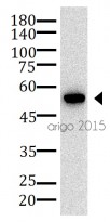
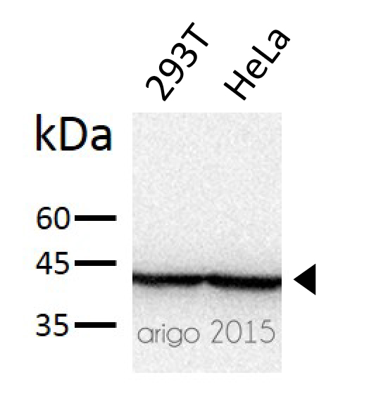
ARG65680 anti-GAPDH antibody WB image
Western blot: 20 µg of 293T and HeLa cell lysate stained with ARG65680 anti-GAPDH antibody at 1:3000 dilution.
-
ARG65693 anti-alpha Tubulin antibody WB image
Western blot: 30 µg of 1) Mouse brain, and 2) Rat brain lysates stained with ARG65693 anti-alpha Tubulin antibody at 1:500 dilution.
-
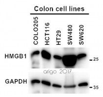
ARG65680 anti-GAPDH antibody WB image
Western blot: 20 µg of COLO205, HCT116, HT29, SW480 and SW620 cell lysates stained with ARG10756 anti-HMGB1 antibody [1F3] (1:2000) and ARG65680 anti-GAPDH antibody (1:10000).
-
ARG65680 anti-GAPDH antibody WB image
Western blot: 30 µg of 1) MDA-MB-231, and 2) MCF-7 lysates stained with ARG65680 anti-GAPDH antibody at 1:5000 dilution.
-
ARG65680 anti-GAPDH antibody WB image
Western blot: A549 cells stained with ARG65680 anti-GAPDH antibody at 1:10000 dilution.
From Youwei Huang et al. Oncol Rep (2022), doi: 10.3892/or.2022.8396, Fig. 4. C.
-
ARG65680 anti-GAPDH antibody WB image
Western blot: Patient-derived GBM cells stained with ARG65680 anti-GAPDH antibody at 1:5000 dilution.
From Xueqin Chen et al. Nat Commun (2023), doi: 10.1038/s41467-023-42545-3, Fig. 7. A.
-

ARG65680 anti-GAPDH antibody WB image
Western blot: CRC cell lines stained with ARG65680 anti-GAPDH antibody at 1:10000 dilution.
From Yuqing Yang et al. Oncol Rep (2024), doi: 10.3892/or.2024.8813, Fig. 4. A.
-

ARG65683 anti-beta Actin antibody WB image
Western blot: HCT116 cells stained with ARG65683 anti-beta Actin antibody.
From Fang Wang et al. Cell Rep (2023), doi: 10.1016/j.celrep.2023.113318, Fig. S2. A.
-

ARG65693 anti-alpha Tubulin antibody WB image
Western blot: A549 cells stained with ARG65693 anti-alpha Tubulin antibody at 1:10000 dilution.
From Youwei Huang et al. Oncol Rep (2022), doi: 10.3892/or.2022.8396, Fig. 4. A.
-

ARG65693 anti-alpha Tubulin antibody WB image
Western blot: HCT116 cells stained with ARG65693 anti-alpha Tubulin antibody.
From Fang Wang et al. Cell Rep (2023), doi: 10.1016/j.celrep.2023.113318, Fig. 3. A.
-
ARG65693 anti-alpha Tubulin antibody WB image
Western blot: Rat sample stained with ARG65693 anti-alpha Tubulin antibody at 1:5000 dilution.
From Jinzhi Li et al. J Reprod Immunol (2023), doi: 10.1016/j.jri.2023.104166, Fig. 2. B.
-

ARG65693 anti-alpha Tubulin antibody WB image
Western blot: Glioma cells stained with ARG65693 anti-alpha Tubulin antibody at 1:5000 dilution.
From Xueqin Chen et al. Nat Commun (2023), doi: 10.1038/s41467-023-42545-3, Fig. 2. D.
-
ARG65693 anti-alpha Tubulin antibody WB image
Western blot: HCT116 and SF763 cells stained with ARG65693 anti-alpha Tubulin antibody at 1:10000 dilution.
From Yuqing Yang et al. Oncol Rep (2024), doi: 10.3892/or.2024.8813, Fig. 6. B.
-

ARG65693 anti-alpha Tubulin antibody WB image
Western blot: GBM cells stained with ARG65693 anti-alpha Tubulin antibody.
From Jialuo Mai et al. Mol Oncol (2019), doi: 10.1002/1878-0261.12525, Fig. 2. D.
-

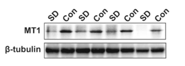
ARG65351 Goat anti-Rabbit IgG antibody (HRP) WB image
Western blot: Rat placental stained with ARG57589 anti-MTNR1A antibody at 1:1000 dilution, ARG65351 Goat anti-Rabbit IgG antibody (HRP) at 1:5000 dilution.
From Jinzhi Li et al. J Reprod Immunol. (2023), doi: 10.1016/j.jri.2023.104166, Fig. 2.B.
-
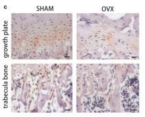
ARG65351 Goat anti-Rabbit IgG antibody (HRP) IHC-P image
From Yu-Qian Song et al. J Mol Med (Berl) (2022), doi: 10.1007/s00109-021-02165-0, Fig. 5.c.


Specific References
Engineering supramolecular dynamics of self-assembly and turnover of oncogenic microRNAs to drive their synergistic destruction in tumor models
ARG65680: WB / Human


Pharmacokinetic enhancement of oncolytic virus M1 by inhibiting JAK‒STAT pathway
ARG65693: WB / Mouse
Arming oncolytic M1 virus with gasdermin E enhances antitumor efficacy in breast cancer
ARG65693; WB / Mouse
Engineered extracellular vesicles with polypeptide for targeted delivery of doxorubicin against EGFR‑positive tumors
ARG65693: WB / Human
Discovery and mechanism of anti-hypertensive effect of a novel tripeptide (SYP) from medicinal fungus Ganoderma lingzhi
ARG65683: WB / Human
A novel function and mechanism of ischemia-induced retinal astrocyte-derived exosomes for RGC apoptosis of ischemic retinopathy
ARG65693: WB / Mouse
Detection of micro-plasma-induced exosomes secretion in a fibroblast-melanoma co-culture model
ARG65693: WB / Mouse
IDH1 mutation impairs antiviral response and potentiates oncolytic virotherapy in glioma
ARG65693: WB /
Sleep deprivation during pregnancy leads to poor fetal outcomes in Sprague-Dawley rats
ARG65693: WB / Rat
RCC2 promotes prostate cancer cell proliferation and migration through Hh/GLI1 signaling pathway and cancer stem-like cells
ARG65680: WB / Human
Targeting VCP potentiates immune checkpoint therapy for colorectal cancer
ARG65693: WB / Mouse
Directly targeting ASC by lonidamine alleviates inflammasome-driven diseases
ARG65693: WB / Mouse
SOX2 regulates paclitaxel resistance of A549 non‑small cell lung cancer cells via promoting transcription of ClC‑3
ARG65680: WB / Human
Fe3O4 Nanozymes Improve Neuroblast Differentiation and Blood-Brain Barrier Integrity of the Hippocampal Dentate Gyrus in D-Galactose-Induced Aged Mice
ARG65683: WB / Mouse
An auto-antibody identified from phenotypic directed screening platform shows host immunity against EV-A71 infection
ARG65683: WB / Human
Tight junction protein 1 promotes vasculature remodeling via regulating USP2/TWIST1 in bladder cancer
ARG65680: WB / Human
Predicting the effects of radiotherapy based on diffusion kurtosis imaging in a xenograft mouse model of esophageal carcinoma
ARG65351: WB /
Design, Synthesis, and Activity Evaluation of Novel Acyclic Nucleosides as Potential Anticancer Agents In Vitro and In Vivo.
ARG65351: WB / Rabbit
Inactivation of the tumor suppressor p53 by long noncoding RNA RMRP.
ARG65351: WB / Rabbit
Intermittent pressure imitating rolling manipulation ameliorates injury in skeletal muscle cells through oxidative stress and lipid metabolism signalling pathways.
ARG65351: WB / Rabbit
The Essential Role of Stathmin in Myoblast C2C12 for Vertical Vibration-Induced Myotube Formation
ARG65683: WB / Rat
Down-regulation of calreticulin promotes apoptosis in hepatic stellate cells.
ARG65351, ARG65683: WB / Human
Identi cation of Potential Novel Pleiotropic Susceptibility Variants Common To Lumbar Spine Bone Mineral Density And Birth Weight.
ARG65351: IHC-P /
Decidual NR2F2-Expressing CD4 + T Cells Promote TH2 Transcriptional Program During Early Pregnancy.
ARG65351: WB /
Baicalin promotes chondrocyte viability and the synthesis of extracellular matrix through TGF-β/Smad3 pathway in chondrocytes
ARG65351: WB / Rabbit
MicroRNA-145 Delay Plaque Erosion by Down-Regulating TGFβr Ii and Suppressing Autophagy of Vascular Smooth Muscle Cells.
ARG65351: WB /
Circ_0081572 inhibits the progression of periodontitis through regulating the miR-378h/RORA axis.
ARG65351: WB /
High expression of FUSE binding protein 1 in breast cancer stimulates cell proliferation and diminishes drug sensitivity.
ARG65351: WB /
Complete zwitterionic double network hydrogels with great toughness and resistance against foreign body reaction and thrombus.
ARG65351: ELISA /
CLC-3 and SOX2 regulate the cell cycle in DU145 cells
ARG65351: WB /
Effect of ERK5 on the expression of iNOS and MCP-1 in M1 type macrophages.
ARG65351: WB /
Ubiquitin ligase DTX3 empowers mutant p53 to promote ovarian cancer development
ARG65351: WB /
Melatonin alleviates progression of uterine endometrial cancer by suppressing estrogen/ubiquitin C/SDHB-mediated succinate accumulation.
ARG65351: WB / Rabbit
BCL7C suppresses ovarian cancer growth by inactivating mutant p53.
ARG65351: WB /
SMARCB1 Promotes Ubiquitination and Degradation of NR4A3 via Direct Interaction Driven by ROS in Vascular Endothelial Cell Injury
ARG65351: WB / Rabbit
The effect of TLR4 on the growth and local inflammatory microenvironment of HPV-related cervical cancer in vivo.
ARG65351: IHC-P /
miR-765 Impairs Pancreatic β-cell Function by Targeting PDX1 in type 2 Diabetes.
ARG65351: WB /
Codelivery of Anti-PD-1 Antibody and Paclitaxel with Matrix Metalloproteinase and pH Dual-Sensitive Micelles for Enhanced Tumor Chemoimmunotherapy.
ARG65351: WB, IHC-P /
4-phenylbutyric acid alleviates bleomycin-induced pulmonary fibrosis in mouse via inhibition of endoplasmic reticulum stress.
ARG65683: WB / Mouse
Baicalin promotes extracellular matrix synthesis in chondrocytes via the activation of hypoxia-inducible factor-1α
ARG65351: WB /
Down-regulation of HTR1A-modulated ACC activation contributes to stress-induced visceral hyperalgesia in rats.
ARG65351: WB / Rabbit
The Different Effects of IFN- β and IFN- γ on the Tumor-Suppressive Activity of Human Amniotic Fluid-Derived Mesenchymal Stem Cells
ARG65683: WB / Human
Ubiquitin ligase TRIM71 suppresses ovarian tumorigenesis by degrading mutant p53.
ARG65351: WB / Rabbit
Icariin suppresses cell cycle transition and cell migration in ovarian cancer cells.
ARG65351: WB / Rabbit
miR-30b-5p acts as a tumor suppressor microRNA in esophageal squamous cell carcinoma.
ARG65680, ARG65351: WB / Human, Rabbit
A Novel Synthetic Steroid of 2β,3α,5α-Trihydroxy-androst-6-one Alleviates the Loss of Rat Retinal Ganglion Cells Caused by Acute Intraocular Hypertension via Inhibiting the Inflammatory Activation of Microglia.
ARG65693: WB / Mouse
Negative Correlation Between Serum Levels of Homocysteine and Apolipoprotein M.
ARG65680: WB /
Alterations ofestradiol-induced histone H3 acetylation in the preoptic area and anteroventral periventricular nucleus of middle-aged female rats.
ARG65351: IHC-P / Rabbit
Negative regulation of miR-1275 by H3K27me3 is critical for glial induction of glioblastoma cells.
ARG65693: WB / Human
Dengue virus nonstructural protein 1 activates platelets via Toll-like receptor 4, leading to thrombocytopenia and hemorrhage.
ARG65683: WB / Human
Icariin increases chondrocyte vitality by promoting hypoxia-inducible factor-1α expression and anaerobic glycolysis.
ARG65351: WB / Rabbit
Treatment of diabetic mice with A Combination of Ketogenic Diet and Aerobic Exercise via modulations of PPARs gene programs.
ARG65351: WB / Rabbit
The antiepileptic drug levetiracetam promotes neuroblast differentiation and expression of superoxide dismutase in the mouse hippocampal dentate gyrus via PI3K/Akt signalling.
ARG65683: WB / Mouse
DNA-PK inhibition synergizes with oncolytic virus M1 by inhibiting antiviral response and potentiating DNA damage.
ARG65693: WB / Human
Short-term urea cycle inhibition in rat liver cells induced by polyethylene glycol
ARG65351: WB / Rabbit
Knockdown of LGALS12 inhibits porcine adipocyte adipogenesis via PKA-Erk1/2 signaling pathway
ARG65351: WB / Rabbit
Effect of palmitic acid on caspase-12 and proliferation and apoptosis of hepatic stellate cells
ARG65351: IHC-P / Rabbit
A novel method of neural differentiation of PC12 cells by using Opti-MEM as a basic induction medium.
ARG65351: WB / Rat
Molecular insights for the biological interactions between polyethylene glycol and cells.
ARG65351: WB / Rabbit
Voluntary wheel exercise alters the levels of miR-494 and miR-696 in the skeletal muscle of C57BL/6 mice.
ARG65351: WB / Rabbit
Topiramate Improves Neuroblast Differentiation of Hippocampal Dentate Gyrus in the D-Galactose-Induced Aging Mice via Its Antioxidant Effects.
ARG65683: WB / Mouse
Study on the inhibition of hyperthermic CO₂ pneumoperitoneum on the proliferation and migration of colon cancer cells and its mechanism.
ARG65351: WB /
1-Methyl-tryptophan attenuates regulatory T cells differentiation due to the inhibition of estrogen-IDO1-MRC2 axis in endometriosis.
ARG65351: WB / Rabbit


