ARG30151
Glucose uptake: Insulin Receptor Dependent Pathway Antibody Panel (GLUT4, AKT pS473, IRS1 pS636)
Cancer antibody; Cell Biology and Cellular Response antibody; Cell Death antibody; Controls and Markers antibody; Developmental Biology antibody; Gene Regulation antibody; Immune System antibody; Metabolism antibody; Neuroscience antibody; Signaling Transduction antibody
Component
| Cat No | Component Name | Host clonality | Reactivity | Application | Package |
|---|---|---|---|---|---|
| ARG51720 | anti-IRS1 phospho (Ser636) antibody | Rabbit pAb | Hu, Ms, Rat | IHC-P, WB | 50 μl |
| ARG51558 | anti-Akt phospho (Ser473) antibody | Rabbit pAb | Hu, Ms, Rat | ICC/IF, IHC-P, WB | 50 μl |
| ARG65292 | anti-GLUT4 antibody | Goat pAb | Hu, Ms, Rat | IHC-P, WB | 50 μg |
| ARG65351 | Goat anti-Rabbit IgG antibody (HRP) | Goat pAb | Rb | ELISA, IHC-P, WB | 50 μl |
| ARG65352 | Donkey anti-Goat IgG antibody (HRP) | Donkey pAb | Goat | ELISA, IHC-P, WB | 50 μl |
Overview
| Product Description | Glucose is the primary energy source for most cells of the body. The study of glucose metabolism is central to cell proliferation, growth, survival, and most recently, tumor progression. Insulin stimulates glucose uptake from blood into skeletal muscle and adipose tissue through a signaling cascade mediated by the insulin receptor (IR). Insulin binding to the IR results in activation of the insulin receptor substrate (IRS) protein and subsequent signaling to the PI3K/Akt pathways, resulting in translocation of Glut4 vesicles, glucose uptake, cell proliferation, and survival. Otherwise, glucose uptake also can be stimulated by adrenoceptor. Upon ligand binding, b3-adrenoceptors result in increased levels of intracellular cAMP that lead to increased de novo synthesis of GLUT1 and mTORC2 phosphorylation. The newly produced GLUT1 is then translocated to the plasma membrane and helps the glucose uptake from blood. These two antibody PANELs can be used to identify the glucose uptake through insulin or adrenoceptors dependent pathway. Mihaylova MM, Shaw RJ (2011) Nat. Cell Biol. 13(9), 1016–23. Zemva J, Schubert M (2011) Curr Diabetes Rev 7(5), 356–66. Olsen, J. M. et al (2014) J. Cell Biol.207(3), 365-374. |
|---|---|
| Target Name | Glucose uptake: Insulin Receptor Dependent Pathway |
| Alternate Names | Glucose uptake: Insulin Receptor Dependent Pathway antibody; Akt phospho (Ser473) antibody; IRS1 phospho (Ser636) antibody; GLUT4 antibody |
Properties
| Storage Instruction | For continuous use, store undiluted antibody at 2-8°C for up to a week. For long-term storage, aliquot and store at -20°C or below. Storage in frost free freezers is not recommended. Avoid repeated freeze/thaw cycles. Suggest spin the vial prior to opening. The antibody solution should be gently mixed before use. |
|---|---|
| Note | For laboratory research only, not for drug, diagnostic or other use. |
Bioinformation
| Gene Full Name | Antibody Panel for Glucose uptake: Insulin Receptor Dependent Pathway (GLUT4, AKT pS473, IRS1 pS636) |
|---|---|
| Highlight | Related Product: anti-GLUT4 antibody; |
| Research Area | Cancer antibody; Cell Biology and Cellular Response antibody; Cell Death antibody; Controls and Markers antibody; Developmental Biology antibody; Gene Regulation antibody; Immune System antibody; Metabolism antibody; Neuroscience antibody; Signaling Transduction antibody |
Images (13) Click the Picture to Zoom In
-
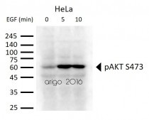
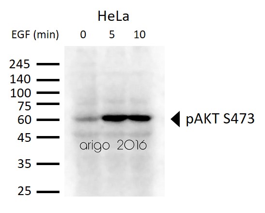
ARG51558 anti-Akt phospho (Ser473) antibody WB image
Western blot: 30 µg of HeLa cells untreated or treated with EGF. Lysates stained with ARG51558 anti-Akt phospho (Ser473) antibody at 1:500 dilution.
-
_210_205.jpg)
ARG65352 Donkey anti-Goat IgG antibody (HRP) WB image
Western blot: Endometrial Stromal cells stained with ARG63922 anti-EP4 prostaglandin Receptor antibody at 1:1000 dilution and ARG65352 Donkey anti-Goat IgG antibody (HRP).
From Qingqing Huang et al. Reprod Med Biol. (2021), doi: 10.1002/rmb2.12423, Fig. 3.C.
-
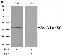
ARG51558 anti-Akt phospho (Ser473) antibody WB image
Western Blot: extracts from 293 cells, treatedwith EGF or calf intestinal phosphatase (CIP), stained with anti-Akt (phospho Ser473) antibody ARG51558.
-
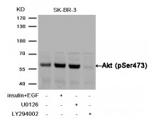
ARG51558 anti-Akt phospho (Ser473) antibody WB image
Western Blot: extracts from SK-BR-3 cells,treated with insulin and EGF, and pretreated with U0126and LY294002 cells stained with anti-Akt (phospho Ser473) antibody ARG51558.
-

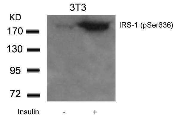
ARG51720 anti-IRS-1 phospho (Ser636) antibody WB image
Western Blot: extracts from 3T3 cells untreated or treated with Insulin stained with anti-IRS-1 (phospho Ser636) antibody ARG51720.
-

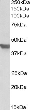
ARG65292 anti-SLC2A4 / GLUT4 antibody WB image
Western Blot: Mouse Heart lysate (35 µg protein in RIPA buffer) stained with ARG65292 anti-SLC2A4 / GLUT4 antibody at 1 µg/ml dilution.
-
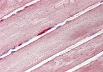
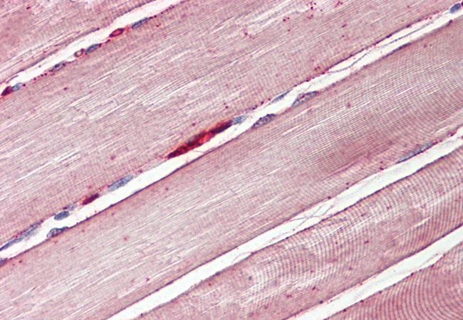
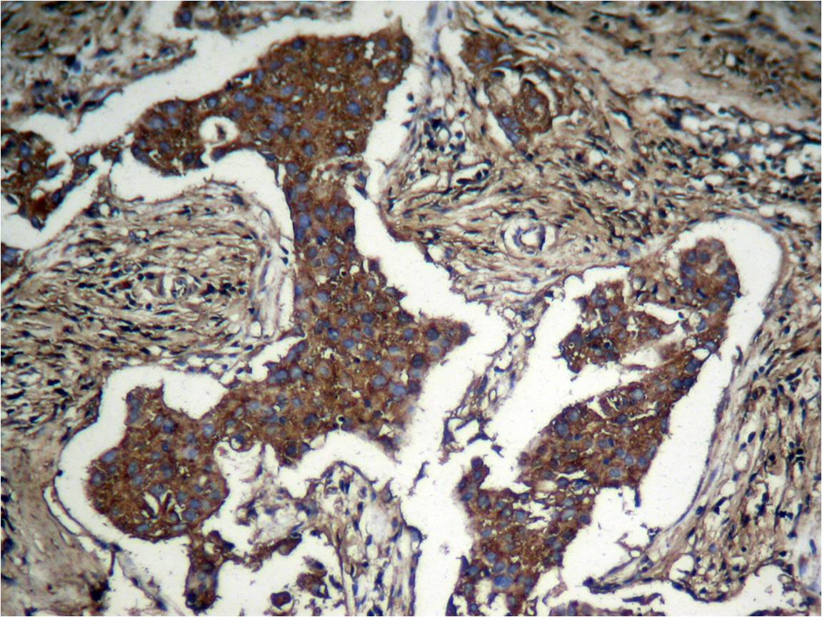
ARG65292 anti-GLUT4 antibody IHC-P image
Immunohistochemistry: Paraffin-embedded Human skeletal muscle tissue. Antigen Retrieval: Steam tissue section in Citrate buffer (pH 6.0). The tissue section was stained with ARG65292 anti-GLUT4 antibody at 3.75 µg/ml dilution followed by AP-staining.
-
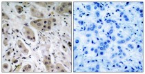
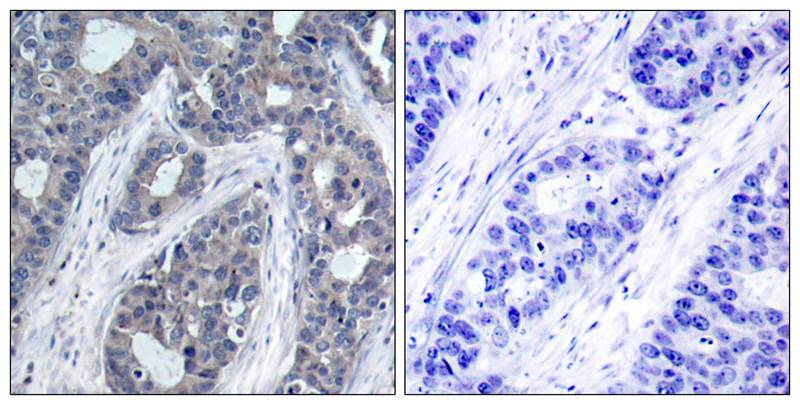
ARG51558 anti-Akt phospho (Ser473) antibody IHC-P image
Immunohistochemistry: paraffin-embedded human breast carcinoma tissue stained with anti-Akt (phospho Ser473) antibody ARG51558 (left) or the same antibody preincubated with blocking peptide (right).
-
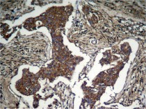
ARG51558 anti-Akt phospho (Ser473) antibody IHC-P image
Immunohistochemistry: paraffinembeddedhuman Lung carcinoma tissue stained with anti-Akt (phospho Ser473) antibody ARG51558.
-

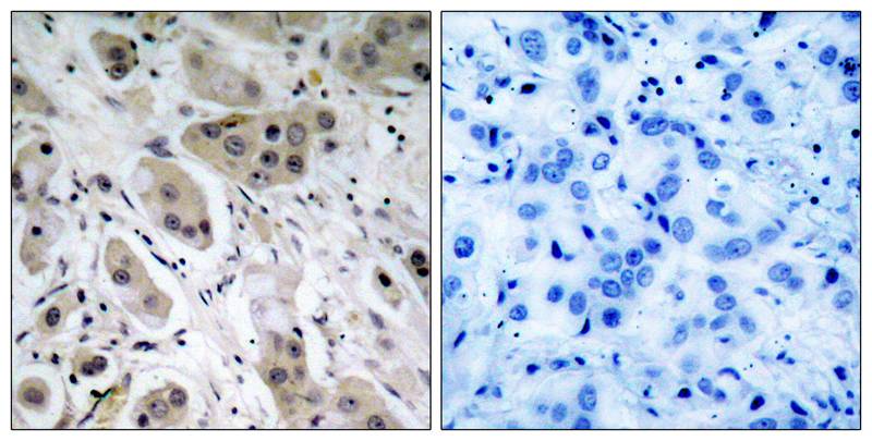
ARG51720 anti-IRS-1 phospho (Ser636) antibody IHC-P image
Immunohistochemistry: paraffin-embedded human breast carcinoma tissue stained with anti-IRS-1 (phospho Ser636) antibody ARG51720 (left) or the same antibody preincubated with blocking peptide (right).
-

ARG65351 Goat anti-Rabbit IgG antibody (HRP) WB image
Western blot: Rat placental stained with ARG57589 anti-MTNR1A antibody at 1:1000 dilution, ARG65351 Goat anti-Rabbit IgG antibody (HRP) at 1:5000 dilution.
From Jinzhi Li et al. J Reprod Immunol. (2023), doi: 10.1016/j.jri.2023.104166, Fig. 2.B.
-

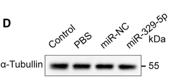
ARG65351 Goat anti-Rabbit IgG antibody (HRP) WB image
Western blot: Mouse retina stained with ARG65693 anti-alpha Tubulin antibody and ARG65351 Goat anti-Rabbit IgG antibody (HRP)
From Xiaoyuan Ye et al. Mol Ther Nucleic Acids. (2024), doi: 10.1016/j.omtn.2024.102209, Fig. 5.D.
-
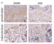
ARG65351 Goat anti-Rabbit IgG antibody (HRP) IHC-P image
From Yu-Qian Song et al. J Mol Med (Berl) (2022), doi: 10.1007/s00109-021-02165-0, Fig. 5.c.
.jpg)


Specific References
The Therapeutic Potential of Exosomes vs. Matrix-Bound Nanovesicles from Human Umbilical Cord Mesenchymal Stromal Cells in Osteoarthritis Treatment
ARG65351; WB /


Environmental acidification drives inter-organ energy mobilization to enhance reproductive performance in medaka (Oryzias latipes)
ARG65351; WB /
Sika Deer Velvet Antler Peptide Exerts Neuroprotective Effect in a Parkinson's Disease Model via Regulating Oxidative Damage and Gut Microbiota
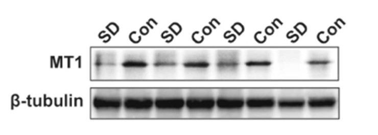
ARG51558: WB / Mouse
Downregulation of AKT/mTOR signaling pathway for Salmonella-mediated autophagy in human anaplastic thyroid cancer
ARG51558: IHC-P / Mouse
Mild hypothermia protects rat cortical neurons against oxygen-glucose deprivation/reoxygenation injury via the PI3K/Akt pathway.
ARG51558: WB, ICC/IF / Rat
Dopamine D2 receptor regulates cortical synaptic pruning in rodents
ARG51558: WB / Rat
1,3-dichloro-2-propanol induced lipid accumulation by blocking autophagy flux in HepG2 cells.
ARG51558: WB / Human
Pharmacological blockage of ICAM-1 improves angiotensin II-induced cardiac remodeling by inhibiting adhesion of LFA-1+ monocytes.
ARG51558: WB / Mouse
Protective effects of dioscin against fructose-induced renal damage via adjusting Sirt3-mediatedoxidative stress, fibrosis, lipid metabolism and inflammation.
ARG51558: WB / Rat
电针对控制性超排卵小鼠子宫内膜磷酸化胰岛素样生长因子-1受体表达及其信号转导通路的调控
ARG51558: WB / Mouse


