ARG30007
Astrocyte Marker Antibody Duo (Host: Goat, Rabbit)
Controls and Markers antibody; Developmental Biology antibody; Neuroscience antibody; Signaling Transduction antibody
Component
| Cat No | Component Name | Host clonality | Reactivity | Application | Package |
|---|---|---|---|---|---|
| ARG52312 | anti-GFAP antibody | Rabbit pAb | Ms, Rat | ICC/IF, IHC-P, IHC-Fr, IHC-FoFr , WB | 50 μl |
| ARG64066 | anti-GFAP antibody | Goat pAb | Hu, Ms, Rat | IHC-Fr, IHC-P, WB | 50 μg |
Overview
| Product Description | Glial fibrillary acidic protein (GFAP) is the major intermediate filament protein in mature astrocytes, a main type of glial cells in the central nervous system (CNS). GFAP is used as a marker to distinguish astrocytes from other glial cells during development. If the CNS is injured through acute CNS trauma, ischemia or neurodegenerative diseases, astrocytes react by rapidly producing more GFAP. Upregulation of GFAP expression has become a pathological hallmark of CNS lesions. arigo's ARG30007 Astrocyte Marker Antibody Duo (Host: Goat, Rabbit) comprises both rabbit and goat anti-GFAP antibodies that are excellent solution for co-staining with other neural lineage marker or brain injury marker. Related news: Astrocyte-to-neuron conversion for Parkinson's disease treatment |
|---|---|
| Target Name | Astrocyte Marker |
| Alternate Names | Astrocyte Marker antibody; GFAP antibody |
Properties
| Storage Instruction | For continuous use, store undiluted antibody at 2-8°C for up to a week. For long-term storage, aliquot and store at -20°C or below. Storage in frost free freezers is not recommended. Avoid repeated freeze/thaw cycles. Suggest spin the vial prior to opening. The antibody solution should be gently mixed before use. |
|---|---|
| Note | For laboratory research only, not for drug, diagnostic or other use. |
Bioinformation
| Gene Full Name | Antibody Duo for Astrocyte Marker |
|---|---|
| Highlight | Related Product: anti-GFAP antibody; |
| Research Area | Controls and Markers antibody; Developmental Biology antibody; Neuroscience antibody; Signaling Transduction antibody |
Images (7) Click the Picture to Zoom In
-
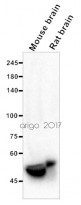
ARG64066 anti-GFAP antibody WB image
Western blot: 20 µg of Mouse brain and Rat brain lysates stained with ARG64066 anti-GFAP antibody at 1:5000 dilution.
-
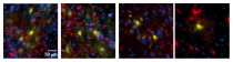
ARG52312 anti-GFAP antibody IHC-Fr image
Immunohistochemistry: Frozen section of Mouse C57BL/6Jnarl brain tissue. The tissue section was fixed by 4% formalin and blocked with BSA with 3% Goat serum, at RT for 1 hour. Tissue section was then stained with ARG52312 anti-GFAP antibody at 1:500 dilution, in PBS with 1% Goat serum, overnight at 4°C.
Blue: DAPI
Yellow: Venus reporter gene
Red: GFAP -
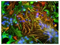
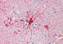
ARG52312 anti-GFAP antibody ICC/IF image
Immunofluorescence: Cultured neurons and glia stained with ARG52312 anti-GFAP antibody (red) and ARG52468 anti-Vimentin antibody (green) showing specific labeling of GFAP (red) and vimentin (green). Cells containing GFAP and vimentin appear yellow
-
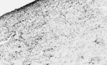
ARG64066 anti-GFAP antibody IHC-Fr image
Immunohistochemistry: PFA-perfused cryosection of Human hypothalamus tissue stained with ARG64066 anti-GFAP antibody at 0.01 µg/ml dilution.
-
ARG64066 anti-GFAP antibody IHC-P image
Immunohistochemistry: Paraffin-embedded Human cortex tissue. Antigen Retrieval: Steam tissue section in Citrate buffer (pH 6.0). The tissue section was stained with ARG64066 anti-GFAP antibody at 5 µg/ml dilution followed by AP-staining.
-
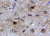
ARG64066 anti-GFAP antibody IHC-P image
Immunohistochemistry: Paraffin-embedded Human cerebellum tissue. Antigen Retrieval: Steam tissue section in Citrate buffer (pH 6.0). The tissue section was stained with ARG64066 anti-GFAP antibody at 2 µg/ml dilution, followed by HRP-staining.
-
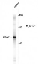
ARG52312 anti-GFAP antibody WB image
Western blot: Rat cortex lysate showing specific immunolabeling of the ~50 kDa GFAP protein stained with ARG52312 anti-GFAP antibody.


Specific References
Acupuncture promotes neurological recovery and regulates lymphatic function after acute inflammatory nerve root injury
ARG64066: IHC-Fr / Human


Paclitaxel induces cognitive impairment via necroptosis, decreased synaptic plasticity and M1 polarisation of microglia
ARG52312: IHC-Fr / Mouse
Toll-Like Receptor 4 Mediates Methamphetamine-Induced Neuroinflammation through Caspase-11 Signaling Pathway in Astrocytes
ARG52312: IHC-P, IHC-Fr / Mouse


