ARG64066
anti-GFAP antibody
anti-GFAP antibody for IHC-Frozen sections,IHC-Formalin-fixed paraffin-embedded sections,Western blot and Human,Mouse,Rat
Controls and Markers antibody; Developmental Biology antibody; Neuroscience antibody; Signaling Transduction antibody; Astrocyte Marker antibody; Astrocyte Maturation Marker antibody; Neuroinflammation antibody; Brain Injury IHC Study antibody

Overview
| Product Description | Goat Polyclonal antibody recognizes Glial Fibrillary Acidic Protein (GFAP) |
|---|---|
| Tested Reactivity | Hu, Ms, Rat |
| Predict Reactivity | Dog |
| Tested Application | IHC-Fr, IHC-P, WB |
| Specificity | GFAP is thought to help to maintain astrocyte mechanical strength, as well as the shape of cells but its exact function remains poorly understood, despite the number of studies using it as a cell marker. |
| Host | Goat |
| Clonality | Polyclonal |
| Isotype | IgG |
| Target Name | GFAP |
| Antigen Species | Human |
| Immunogen | C-DGEVIKESKQEHKD |
| Conjugation | Un-conjugated |
| Alternate Names | Glial fibrillary acidic protein; ALXDRD; GFAP |
Application Instructions
| Application Suggestion |
|
||||||||
|---|---|---|---|---|---|---|---|---|---|
| Application Note | WB: Recommend incubate at RT for 1h. IHC-P: Antigen Retrieval: Steam tissue section in Citrate buffer (pH 6.0). * The dilutions indicate recommended starting dilutions and the optimal dilutions or concentrations should be determined by the scientist. |
Properties
| Form | Liquid |
|---|---|
| Purification | Purified from goat serum by antigen affinity chromatography. |
| Buffer | Tris saline (pH 7.3), 0.02% Sodium azide and 0.5% BSA. |
| Preservative | 0.02% Sodium azide |
| Stabilizer | 0.5% BSA |
| Concentration | 0.5 mg/ml |
| Storage Instruction | For continuous use, store undiluted antibody at 2-8°C for up to a week. For long-term storage, aliquot and store at -20°C or below. Storage in frost free freezers is not recommended. Avoid repeated freeze/thaw cycles. Suggest spin the vial prior to opening. The antibody solution should be gently mixed before use. |
| Note | For laboratory research only, not for drug, diagnostic or other use. |
Bioinformation
| Database Links | |
|---|---|
| Gene Full Name | glial fibrillary acidic protein |
| Background | GFAP is one of the major intermediate filament proteins of mature astrocytes. It is used as a marker to distinguish astrocytes from other glial cells during development. Mutations in this gene cause Alexander disease, a rare disorder of astrocytes in the central nervous system. Alternative splicing results in multiple transcript variants encoding distinct isoforms. [provided by RefSeq, Oct 2008] |
| Function | GFAP is a class-III intermediate filament. It is a cell-specific marker that, during the development of the central nervous system, distinguishes astrocytes from other glial cells. [UniProt] |
| Highlight | Related Antibody Duos and Panels: ARG30007 Astrocyte Marker Antibody Duo (Host: Goat, Rabbit) Related products: GFAP antibodies; GFAP Duos / Panels; Anti-Goat IgG secondary antibodies; Related news: Astrocyte-to-neuron conversion for Parkinson's disease treatment |
| Research Area | Controls and Markers antibody; Developmental Biology antibody; Neuroscience antibody; Signaling Transduction antibody; Astrocyte Marker antibody; Astrocyte Maturation Marker antibody; Neuroinflammation antibody; Brain Injury IHC Study antibody |
| Calculated MW | 50 kDa |
| PTM | Phosphorylated by PKN1. |
Images (4) Click the Picture to Zoom In
-
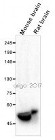
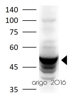
ARG64066 anti-GFAP antibody WB image
Western blot: 20 µg of Mouse brain and Rat brain lysates stained with ARG64066 anti-GFAP antibody at 1:5000 dilution.
-
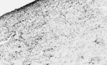
ARG64066 anti-GFAP antibody IHC-Fr image
Immunohistochemistry: PFA-perfused cryosection of Human hypothalamus tissue stained with ARG64066 anti-GFAP antibody at 0.01 µg/ml dilution.
-
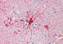
ARG64066 anti-GFAP antibody IHC-P image
Immunohistochemistry: Paraffin-embedded Human cortex tissue. Antigen Retrieval: Steam tissue section in Citrate buffer (pH 6.0). The tissue section was stained with ARG64066 anti-GFAP antibody at 5 µg/ml dilution followed by AP-staining.
-
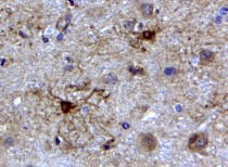
ARG64066 anti-GFAP antibody IHC-P image
Immunohistochemistry: Paraffin-embedded Human cerebellum tissue. Antigen Retrieval: Steam tissue section in Citrate buffer (pH 6.0). The tissue section was stained with ARG64066 anti-GFAP antibody at 2 µg/ml dilution, followed by HRP-staining.


Customer's Feedback
Excellent

Review for anti-GFAP antibody
Application:WB
Sample:Mouse brain and Rat brain
Sample Loading Amount:20 µg
Primary Antibody Dilution Factor:1:5000
Primary Antibody Incubation Time:overnight
Primary Antibody Incubation Temperature:4 ºC

Excellent
Review for anti-GFAP antibody
Application:WB
Sample:Rat brain
Sample Loading Amount:30 µg
Primary Antibody Dilution Factor:1:800
Primary Antibody Incubation Time:overnight
Primary Antibody Incubation Temperature:4 ºC
Specific References
Acupuncture promotes neurological recovery and regulates lymphatic function after acute inflammatory nerve root injury
IHC-Fr / Human


