ARG58206
anti-p21 antibody [DCS-60.2]
anti-p21 antibody [DCS-60.2] for Flow cytometry,ICC/IF,IHC-Formalin-fixed paraffin-embedded sections,Western blot and Human
Overview
| Product Description | Mouse Monoclonal antibody [DCS-60.2] recognizes p21 |
|---|---|
| Tested Reactivity | Hu |
| Species Does Not React With | Ms, Rat |
| Tested Application | FACS, ICC/IF, IHC-P, WB |
| Host | Mouse |
| Clonality | Monoclonal |
| Clone | DCS-60.2 |
| Isotype | IgG2a, kappa |
| Target Name | p21 |
| Antigen Species | Human |
| Immunogen | Recombinant Human p21 protein. |
| Conjugation | Un-conjugated |
| Alternate Names | Melanoma differentiation-associated protein 6; WAF1; CIP1; CDKN1; CAP20; MDA-6; SDI1; CDK-interacting protein 1; P21; p21CIP1; p21; Cyclin-dependent kinase inhibitor 1 |
Application Instructions
| Application Suggestion |
|
||||||||||
|---|---|---|---|---|---|---|---|---|---|---|---|
| Application Note | IHC-P: Antigen Retrieval: Boil tissue section in 10 mM Citrate buffer (pH 6.0) for 10-20 min, followed by cooling at RT for 20 min. * The dilutions indicate recommended starting dilutions and the optimal dilutions or concentrations should be determined by the scientist. |
Properties
| Form | Liquid |
|---|---|
| Purification | Purification with Protein G. |
| Buffer | PBS, 0.05% Sodium azide and 0.1 mg/ml BSA. |
| Preservative | 0.05% Sodium azide |
| Stabilizer | 0.1 mg/ml BSA |
| Concentration | 0.2 mg/ml |
| Storage Instruction | For continuous use, store undiluted antibody at 2-8°C for up to a week. For long-term storage, aliquot and store at -20°C or below. Storage in frost free freezers is not recommended. Avoid repeated freeze/thaw cycles. Suggest spin the vial prior to opening. The antibody solution should be gently mixed before use. |
| Note | For laboratory research only, not for drug, diagnostic or other use. |
Bioinformation
| Database Links |
Swiss-port # P38936 Human Cyclin-dependent kinase inhibitor 1 |
|---|---|
| Gene Symbol | CDKN1A |
| Gene Full Name | cyclin-dependent kinase inhibitor 1A (p21, Cip1) |
| Background | This gene encodes a potent cyclin-dependent kinase inhibitor. The encoded protein binds to and inhibits the activity of cyclin-cyclin-dependent kinase2 or -cyclin-dependent kinase4 complexes, and thus functions as a regulator of cell cycle progression at G1. The expression of this gene is tightly controlled by the tumor suppressor protein p53, through which this protein mediates the p53-dependent cell cycle G1 phase arrest in response to a variety of stress stimuli. This protein can interact with proliferating cell nuclear antigen, a DNA polymerase accessory factor, and plays a regulatory role in S phase DNA replication and DNA damage repair. This protein was reported to be specifically cleaved by CASP3-like caspases, which thus leads to a dramatic activation of cyclin-dependent kinase2, and may be instrumental in the execution of apoptosis following caspase activation. Mice that lack this gene have the ability to regenerate damaged or missing tissue. Multiple alternatively spliced variants have been found for this gene. [provided by RefSeq, Sep 2015] |
| Function | May be the important intermediate by which p53/TP53 mediates its role as an inhibitor of cellular proliferation in response to DNA damage. Binds to and inhibits cyclin-dependent kinase activity, preventing phosphorylation of critical cyclin-dependent kinase substrates and blocking cell cycle progression. Functions in the nuclear localization and assembly of cyclin D-CDK4 complex and promotes its kinase activity towards RB1. At higher stoichiometric ratios, inhibits the kinase activity of the cyclin D-CDK4 complex. [UniProt] |
| Cellular Localization | Nuclear. [UniProt] |
| Highlight | Related products: p21 antibodies; p21 Duos / Panels; Anti-Mouse IgG secondary antibodies; Related news: Senescence Marker Antibody Panel is launched |
| Calculated MW | 18 kDa |
| PTM | Phosphorylation of Thr-145 by Akt or of Ser-146 by PKC impairs binding to PCNA. Phosphorylation at Ser-114 by GSK3-beta enhances ubiquitination by the DCX(DTL) complex. Phosphorylation of Thr-145 by PIM2 enhances CDKN1A stability and inhibits cell proliferation. Phosphorylation of Thr-145 by PIM1 results in the relocation of CDKN1A to the cytoplasm and enhanced CDKN1A protein stability. UV radiation-induced phosphorylation at Thr-80 by LKB1 and at Ser-146 by NUAK1 leads to its degradation. Ubiquitinated by MKRN1; leading to polyubiquitination and 26S proteasome-dependent degradation. Ubiquitinated by the DCX(DTL) complex, also named CRL4(CDT2) complex, leading to its degradation during S phase or following UV irradiation. Ubiquitination by the DCX(DTL) complex is essential to control replication licensing and is PCNA-dependent: interacts with PCNA via its PIP-box, while the presence of the containing the 'K+4' motif in the PIP box, recruit the DCX(DTL) complex, leading to its degradation. Ubiquitination at Ser-2 leads to degradation by the proteasome pathway. Ubiquitinated by RNF114; leading to proteasomal degradation. Acetylation leads to protein stability. Acetylated in vitro on Lys-141, Lys-154, Lys-161 and Lys-163. Deacetylation by HDAC1 is prevented by competitive binding of C10orf90/FATS to HDAC1 (By similarity). [UniProt] |
Images (2) Click the Picture to Zoom In
-
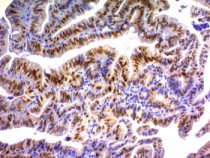
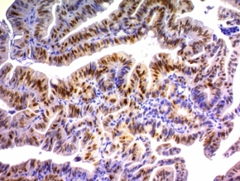
ARG58206 anti-p21 antibody [DCS-60.2] IHC-P image
Immunohistochemistry: Formalin-fixed and paraffin-embedded Human colon carcinoma stained with ARG58206 anti-p21 antibody [DCS-60.2].
-
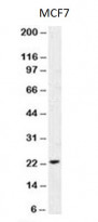
ARG58206 anti-p21 antibody [DCS-60.2] WB image
Western blot: MCF7 cell lysate stained with ARG58206 anti-p21 antibody [DCS-60.2].