ARG63119
anti-Vimentin antibody [VI-RE/1]
anti-Vimentin antibody [VI-RE/1] for ELISA,Flow cytometry,ICC/IF,IHC-Formalin-fixed paraffin-embedded sections,Western blot and Human
Cancer antibody; Controls and Markers antibody; Developmental Biology antibody; Neuroscience antibody; Signaling Transduction antibody; Cancer-associated fibroblast antibody; CAF Marker antibody; EMT Study antibody; Mesenchymal Markers antibody; Fibroblast Marker antibody; Muller Cell Marker antibody; Sarcoma Marker antibody
Overview
| Product Description | Mouse Monoclonal antibody [VI-RE/1] recognizes Vimentin |
|---|---|
| Tested Reactivity | Hu |
| Species Does Not React With | Ms, Pig |
| Tested Application | ELISA, FACS, ICC/IF, IHC-P, WB |
| Specificity | The clone VI-RE/1 reacts with human vimentin, a 57 kDa intermediate filament protein expressed on a wide variety of mesenchymal and mesodermal cell types. |
| Host | Mouse |
| Clonality | Monoclonal |
| Clone | VI-RE/1 |
| Isotype | IgG1 |
| Target Name | Vimentin |
| Antigen Species | Human |
| Immunogen | Bacterially expressed full-length human vimentin |
| Conjugation | Un-conjugated |
| Alternate Names | Vimentin; CTRCT30; HEL113 |
Application Instructions
| Application Suggestion |
|
||||||||||||
|---|---|---|---|---|---|---|---|---|---|---|---|---|---|
| Application Note | WB: Incubation: Overnight at 4°C. WB: Sample preparation: Resuspend approx. 50 mil. cells in 1 ml cold Lysis buffer (1% laurylmaltoside in 20 mM Tris/Cl, 100 mM NaCl pH 8.2, 50 mM NaF including Protease inhibitor Cocktail). Incubate 60 min on ice. Centrifuge to remove cell debris. Mix lysate with reducing Laemmli SDS-PAGE sample buffer. Boil for 3 min in water bath. Application note: Reducing condition. SDS-PAGE (10% separating gel). * The dilutions indicate recommended starting dilutions and the optimal dilutions or concentrations should be determined by the scientist. |
||||||||||||
| Positive Control | U87 (positive control); Raji (negative control); MOLT-4 (low expression) | ||||||||||||
| Observed Size | ~ 55 kDa |
Properties
| Form | Liquid |
|---|---|
| Purification | Purified from hybridoma culture supernatant by protein-A affinity chromatography. |
| Purity | > 95% (by SDS-PAGE) |
| Buffer | PBS (pH 7.4) and 15 mM Sodium azide |
| Preservative | 15 mM Sodium azide |
| Concentration | 1 mg/ml |
| Storage Instruction | For continuous use, store undiluted antibody at 2-8°C for up to a week. For long-term storage, aliquot and store at -20°C or below. Storage in frost free freezers is not recommended. Avoid repeated freeze/thaw cycles. Suggest spin the vial prior to opening. The antibody solution should be gently mixed before use. |
| Note | For laboratory research only, not for drug, diagnostic or other use. |
Bioinformation
| Database Links | |
|---|---|
| Gene Symbol | VIM |
| Gene Full Name | vimentin |
| Background | Vimentin is a type III intermediate filament protein. Intermediate filaments, along with microtubules and actin microfilaments, make up the cytoskeleton. The encoded protein is responsible for maintaining cell shape and integrity of the cytoplasm, and stabilizing cytoskeletal interactions. This protein is involved in neuritogenesis and cholesterol transport and functions as an organizer of a number of other critical proteins involved in cell attachment, migration, and signaling. Bacterial and viral pathogens have been shown to attach to this protein on the host cell surface. Mutations in this gene are associated with congenital cataracts in human patients. [provided by RefSeq, Aug 2017] |
| Function | Vimentins are class-III intermediate filaments found in various non-epithelial cells, especially mesenchymal cells. Vimentin is attached to the nucleus, endoplasmic reticulum, and mitochondria, either laterally or terminally. Involved with LARP6 in the stabilization of type I collagen mRNAs for CO1A1 and CO1A2. [UniProt] |
| Highlight | Related products: Vimentin antibodies; Vimentin Duos / Panels; Anti-Mouse IgG secondary antibodies; Related news: New antibody panels for Myofibroblasts and CAFs New antibody panels and duos for Tumor immune microenvironment Anti-SerpinB9 therapy, a new strategy for cancer therapy |
| Research Area | Cancer antibody; Controls and Markers antibody; Developmental Biology antibody; Neuroscience antibody; Signaling Transduction antibody; Cancer-associated fibroblast antibody; CAF Marker antibody; EMT Study antibody; Mesenchymal Markers antibody; Fibroblast Marker antibody; Muller Cell Marker antibody; Sarcoma Marker antibody |
| Calculated MW | 54 kDa |
| PTM | Filament disassembly during mitosis is promoted by phosphorylation at Ser-55 as well as by nestin (By similarity). One of the most prominent phosphoproteins in various cells of mesenchymal origin. Phosphorylation is enhanced during cell division, at which time vimentin filaments are significantly reorganized. Phosphorylation by PKN1 inhibits the formation of filaments. Phosphorylated at Ser-56 by CDK5 during neutrophil secretion in the cytoplasm. Phosphorylated by STK33. O-glycosylated during cytokinesis at sites identical or close to phosphorylation sites, this interferes with the phosphorylation status. S-nitrosylation is induced by interferon-gamma and oxidatively-modified low-densitity lipoprotein (LDL(ox)) possibly implicating the iNOS-S100A8/9 transnitrosylase complex. |
Images (3) Click the Picture to Zoom In
-

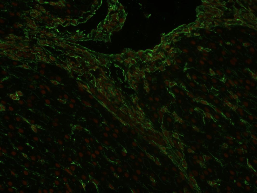
ARG63119 anti-Vimentin antibody [VI-RE/1] IHC-P image
Immunohistochemistry: Paraffin-embedded Human liver tissue stained with ARG63119 anti-Vimentin antibody [VI-RE/1] (green) at 1:400 dilution. Cell nuclei stained with PI (orange) at 1 µg/ml dilution.
-
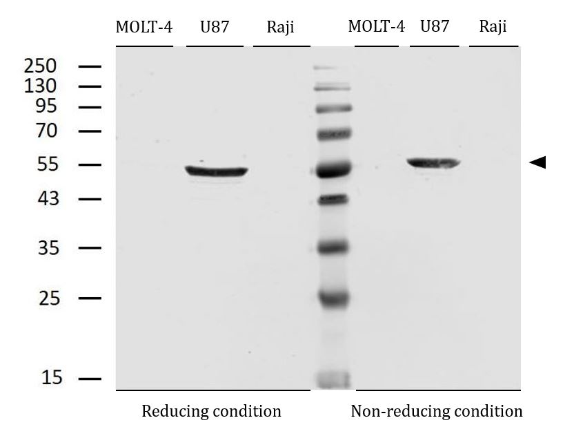
ARG63119 anti-Vimentin antibody [VI-RE/1] WB image
Western blot: MOLT-4 (low expression), U87 (positive) and Raji (negative control) under reducing and non-reducing conditions. The plots were stained with ARG63119 anti-Vimentin antibody [VI-RE/1] at 2 µg/ml dilution.
-

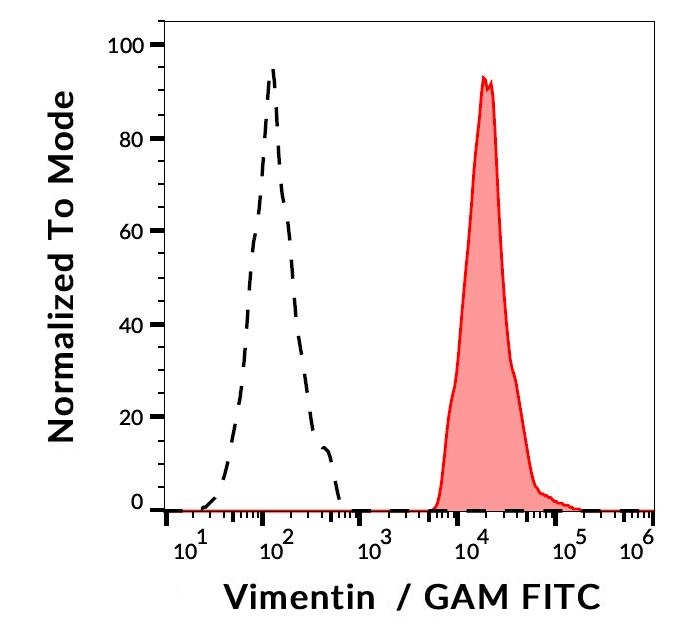
ARG63119 anti-Vimentin antibody [VI-RE/1] FACS image
Flow Cytometry: ESS-1 cells (red) and Human lymphocytes (black-dashed, negative control) stained with ARG63119 anti-Vimentin antibody [VI-RE/1], followed by incubation with FITC labelled Goat anti-Mouse secondary antibody.