ARG54309
anti-Vimentin antibody [VI-RE/1] (APC)
anti-Vimentin antibody [VI-RE/1] (APC) for Flow cytometry and Human
Cancer antibody; Controls and Markers antibody; Developmental Biology antibody; Neuroscience antibody; Signaling Transduction antibody; Cancer-associated fibroblast antibody; CAF Marker antibody; EMT Study antibody; Mesenchymal Markers antibody; Fibroblast Marker antibody; Muller Cell Marker antibody; Sarcoma Marker antibody
Overview
| Product Description | APC-conjugated Mouse Monoclonal antibody [VI-RE/1] recognizes Vimentin |
|---|---|
| Tested Reactivity | Hu |
| Species Does Not React With | Ms, Pig |
| Tested Application | FACS |
| Specificity | The clone VI-RE/1 reacts with human vimentin, a 57 kDa intermediate filament protein expressed on a wide variety of mesenchymal and mesodermal cell types. |
| Host | Mouse |
| Clonality | Monoclonal |
| Clone | VI-RE/1 |
| Isotype | IgG1 |
| Target Name | Vimentin |
| Antigen Species | Human |
| Immunogen | Bacterially expressed full-length human vimentin |
| Conjugation | APC |
| Alternate Names | Vimentin; CTRCT30; HEL113 |
Application Instructions
| Application Suggestion |
|
||||
|---|---|---|---|---|---|
| Application Note | * The dilutions indicate recommended starting dilutions and the optimal dilutions or concentrations should be determined by the scientist. |
Properties
| Form | Liquid |
|---|---|
| Purification Note | The purified antibody is conjugated with cross-linked Allophycocyanin (APC) under optimum conditions. The conjugate is purified by size-exclusion chromatography. |
| Buffer | PBS, 15 mM Sodium azide and 0.2% (w/v) high-grade protease free BSA |
| Preservative | 15 mM Sodium azide |
| Stabilizer | 0.2% (w/v) high-grade protease free BSA |
| Concentration | 0.1 mg/ml |
| Storage Instruction | Aliquot and store in the dark at 2-8°C. Keep protected from prolonged exposure to light. Avoid repeated freeze/thaw cycles. Suggest spin the vial prior to opening. The antibody solution should be gently mixed before use. |
| Note | For laboratory research only, not for drug, diagnostic or other use. |
Bioinformation
| Database Links | |
|---|---|
| Gene Symbol | VIM |
| Gene Full Name | vimentin |
| Background | Vimentin is a type III intermediate filament protein. Intermediate filaments, along with microtubules and actin microfilaments, make up the cytoskeleton. The encoded protein is responsible for maintaining cell shape and integrity of the cytoplasm, and stabilizing cytoskeletal interactions. This protein is involved in neuritogenesis and cholesterol transport and functions as an organizer of a number of other critical proteins involved in cell attachment, migration, and signaling. Bacterial and viral pathogens have been shown to attach to this protein on the host cell surface. Mutations in this gene are associated with congenital cataracts in human patients. [provided by RefSeq, Aug 2017] |
| Function | Vimentins are class-III intermediate filaments found in various non-epithelial cells, especially mesenchymal cells. Vimentin is attached to the nucleus, endoplasmic reticulum, and mitochondria, either laterally or terminally. Involved with LARP6 in the stabilization of type I collagen mRNAs for CO1A1 and CO1A2. [UniProt] |
| Highlight | Related products: Vimentin antibodies; Vimentin Duos / Panels; Anti-Mouse IgG secondary antibodies; Related news: New antibody panels for Myofibroblasts and CAFs New antibody panels and duos for Tumor immune microenvironment Anti-SerpinB9 therapy, a new strategy for cancer therapy |
| Research Area | Cancer antibody; Controls and Markers antibody; Developmental Biology antibody; Neuroscience antibody; Signaling Transduction antibody; Cancer-associated fibroblast antibody; CAF Marker antibody; EMT Study antibody; Mesenchymal Markers antibody; Fibroblast Marker antibody; Muller Cell Marker antibody; Sarcoma Marker antibody |
| Calculated MW | 54 kDa |
| PTM | Filament disassembly during mitosis is promoted by phosphorylation at Ser-55 as well as by nestin (By similarity). One of the most prominent phosphoproteins in various cells of mesenchymal origin. Phosphorylation is enhanced during cell division, at which time vimentin filaments are significantly reorganized. Phosphorylation by PKN1 inhibits the formation of filaments. Phosphorylated at Ser-56 by CDK5 during neutrophil secretion in the cytoplasm. Phosphorylated by STK33. O-glycosylated during cytokinesis at sites identical or close to phosphorylation sites, this interferes with the phosphorylation status. S-nitrosylation is induced by interferon-gamma and oxidatively-modified low-densitity lipoprotein (LDL(ox)) possibly implicating the iNOS-S100A8/9 transnitrosylase complex. |
Images (1) Click the Picture to Zoom In
-
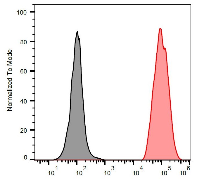
ARG54309 anti-Vimentin antibody [VI-RE/1] (APC) FACS image
Flow Cytometry: Separation of stained ESS-1 cells (red) from unstained ESS-1 cells (black). Cells were stained with ARG54309 anti-Vimentin antibody [VI-RE/1] (APC) at 1 µg/ml dilution.