ARG52469
anti-Vimentin antibody [2D1]
anti-Vimentin antibody [2D1] for ICC/IF,IHC-Frozen sections,Western blot and Human,Mouse,Rat,Bovine,Monkey,Pig
Cancer antibody; Controls and Markers antibody; Developmental Biology antibody; Neuroscience antibody; Signaling Transduction antibody; Cancer-associated fibroblast antibody; CAF Marker antibody; EMT Study antibody; Mesenchymal Markers antibody; Fibroblast Marker antibody; Muller Cell Marker antibody; Sarcoma Marker antibody

Overview
| Product Description | Mouse Monoclonal antibody [2D1] recognizes Vimentin |
|---|---|
| Tested Reactivity | Hu, Ms, Rat, Bov, Mk, Pig |
| Tested Application | ICC/IF, IHC-Fr, WB |
| Host | Mouse |
| Clonality | Monoclonal |
| Clone | 2D1 |
| Isotype | IgG2a |
| Target Name | Vimentin |
| Antigen Species | Human |
| Immunogen | Recombinant human vimentin purified from E. coli |
| Conjugation | Un-conjugated |
| Alternate Names | Vimentin; CTRCT30; HEL113 |
Application Instructions
| Application Suggestion |
|
||||||||
|---|---|---|---|---|---|---|---|---|---|
| Application Note | Specific for the ~50kDa vimentin protein. * The dilutions indicate recommended starting dilutions and the optimal dilutions or concentrations should be determined by the scientist. |
Properties
| Form | Liquid |
|---|---|
| Purification | Affinity Purified |
| Buffer | PBS and 10 mM Sodium azide |
| Preservative | 10 mM Sodium azide |
| Concentration | 1 mg/ml |
| Storage Instruction | For continuous use, store undiluted antibody at 2-8°C for up to a week. For long-term storage, aliquot and store at -20°C or below. Storage in frost free freezers is not recommended. Avoid repeated freeze/thaw cycles. Suggest spin the vial prior to opening. The antibody solution should be gently mixed before use. |
| Note | For laboratory research only, not for drug, diagnostic or other use. |
Bioinformation
| Database Links | |
|---|---|
| Gene Symbol | VIM |
| Gene Full Name | vimentin |
| Background | Vimentin is a type III intermediate filament protein. Intermediate filaments, along with microtubules and actin microfilaments, make up the cytoskeleton. The encoded protein is responsible for maintaining cell shape and integrity of the cytoplasm, and stabilizing cytoskeletal interactions. This protein is involved in neuritogenesis and cholesterol transport and functions as an organizer of a number of other critical proteins involved in cell attachment, migration, and signaling. Bacterial and viral pathogens have been shown to attach to this protein on the host cell surface. Mutations in this gene are associated with congenital cataracts in human patients. [provided by RefSeq, Aug 2017] |
| Function | Vimentins are class-III intermediate filaments found in various non-epithelial cells, especially mesenchymal cells. Vimentin is attached to the nucleus, endoplasmic reticulum, and mitochondria, either laterally or terminally. Involved with LARP6 in the stabilization of type I collagen mRNAs for CO1A1 and CO1A2. [UniProt] |
| Highlight | Related products: Vimentin antibodies; Vimentin Duos / Panels; Anti-Mouse IgG secondary antibodies; Related news: New antibody panels for Myofibroblasts and CAFs New antibody panels and duos for Tumor immune microenvironment Anti-SerpinB9 therapy, a new strategy for cancer therapy |
| Research Area | Cancer antibody; Controls and Markers antibody; Developmental Biology antibody; Neuroscience antibody; Signaling Transduction antibody; Cancer-associated fibroblast antibody; CAF Marker antibody; EMT Study antibody; Mesenchymal Markers antibody; Fibroblast Marker antibody; Muller Cell Marker antibody; Sarcoma Marker antibody |
| Calculated MW | 54 kDa |
| PTM | Filament disassembly during mitosis is promoted by phosphorylation at Ser-55 as well as by nestin (By similarity). One of the most prominent phosphoproteins in various cells of mesenchymal origin. Phosphorylation is enhanced during cell division, at which time vimentin filaments are significantly reorganized. Phosphorylation by PKN1 inhibits the formation of filaments. Phosphorylated at Ser-56 by CDK5 during neutrophil secretion in the cytoplasm. Phosphorylated by STK33. O-glycosylated during cytokinesis at sites identical or close to phosphorylation sites, this interferes with the phosphorylation status. S-nitrosylation is induced by interferon-gamma and oxidatively-modified low-densitity lipoprotein (LDL(ox)) possibly implicating the iNOS-S100A8/9 transnitrosylase complex. |
Images (4) Click the Picture to Zoom In
-

ARG52469 anti-Vimentin antibody [2D1] ICC/IF image
Immunofluorescence: Cortical neuron-glial cell cultures from E20 Rat stained with ARG52469 anti-Vimentin antibody [2D1] (red) at 1:2000 dilution, and costained with anti-GFAP antibody (green) at 1:5000 dilution. DAPI (blue) for nuclear staining.
Fibroblastic and other developing cells express only Vimentin and appear red. Astrocytes that express GFAP are green while those that express both GFAP and Vimentin appear golden yellow.
-
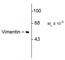
ARG52469 anti-Vimentin antibody [2D1] WB image
Western blot: HeLa cells stained with ARG52469 anti-Vimentin antibody [2D1] showing specific immunolabeling of the ~50k vimentin protein.
-
ARG52469 anti-Vimentin antibody [2D1] ICC/IF image
Immunofluorescence: Mixed neuron/glial cultures stained with anti-vimentin (green) and rabbit anti-GFAP antibody (ARG52312) (red).
Vimentin is expressed alone in fibroblastic and endothelial cells, which are the flattened cells in the middle of the image which appear green.
Astrocytes may express primarily GFAP, or GFAP and vimentin, and so appear red (GFAP only) or golden yellow (GFAP and Vimentin).
In cells which express both GFAP and vimentin, the two proteins assemble to produce heteropolymer filaments. -
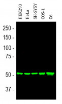
ARG52469 anti-Vimentin antibody [2D1] WB image
Western blot: HEK293, HeLa, SH-SY5Y, COS-1 and C6 cell lysates stained with ARG52469 anti-Vimentin antibody [2D1] (green) at 1:10000 dilution.


Customer's Feedback
Excellent

Review for anti-Vimentin antibody [2D1]
Application:WB
Sample:mda-mB 231 (+ve)
Sample Loading Amount:30 µg
Primary Antibody Dilution Factor:1:1000
Primary Antibody Incubation Time:overnight
Primary Antibody Incubation Temperature:4 ºC


