ARG56882
anti-TP53BP1 antibody
anti-TP53BP1 antibody for IHC-Formalin-fixed paraffin-embedded sections,Western blot and Human,Mouse,Rat

Overview
| Product Description | Rabbit Polyclonal antibody recognizes TP53BP1 |
|---|---|
| Tested Reactivity | Hu, Ms, Rat |
| Tested Application | IHC-P, WB |
| Host | Rabbit |
| Clonality | Polyclonal |
| Isotype | IgG |
| Target Name | TP53BP1 |
| Antigen Species | Human |
| Immunogen | Recombinant protein of Human TP53BP1. |
| Conjugation | Un-conjugated |
| Alternate Names | p53BP1; 53BP1; p202; p53-binding protein 1; Tumor suppressor p53-binding protein 1 |
Application Instructions
| Application Suggestion |
|
||||||
|---|---|---|---|---|---|---|---|
| Application Note | * The dilutions indicate recommended starting dilutions and the optimal dilutions or concentrations should be determined by the scientist. | ||||||
| Positive Control | A431 |
Properties
| Form | Liquid |
|---|---|
| Purification | Affinity purification with immunogen. |
| Buffer | PBS (pH 7.3), 0.02% Sodium azide and 50% Glycerol. |
| Preservative | 0.02% Sodium azide |
| Stabilizer | 50% Glycerol |
| Storage Instruction | For continuous use, store undiluted antibody at 2-8°C for up to a week. For long-term storage, aliquot and store at -20°C. Storage in frost free freezers is not recommended. Avoid repeated freeze/thaw cycles. Suggest spin the vial prior to opening. The antibody solution should be gently mixed before use. |
| Note | For laboratory research only, not for drug, diagnostic or other use. |
Bioinformation
| Database Links |
Swiss-port # Q12888 Human Tumor suppressor p53-binding protein 1 |
|---|---|
| Gene Symbol | TP53BP1 |
| Gene Full Name | tumor protein p53 binding protein 1 |
| Function | Plays a key role in the response to DNA damage. May have a role in checkpoint signaling during mitosis. Enhances TP53-mediated transcriptional activation. [UniProt] |
| Highlight | Related products: TP53BP1 antibodies; Anti-Rabbit IgG secondary antibodies; Related news: Senescence Marker Antibody Panel is launched |
| Calculated MW | 214 kDa |
| PTM | Asymmetrically dimethylated on Arg residues by PRMT1. Methylation is required for DNA binding. Phosphorylated at basal level in the absence of DNA damage (PubMed:11042216, PubMed:11331310). Phosphorylated by ATM in response to DNA damage: phosphorylation at different sites promotes interaction with different set of proteins: phosphorylation at the N-terminus by ATM (residues from 6-178) promotes interaction with PAXIP1 and non-homologous end joining (NHEJ) of dysfunctional telomeres (PubMed:23727112). Phosphorylation by ATM at residues that are located more C-terminus (residues 300-650) leads to promote interaction with RIF1 (PubMed:23727112, PubMed:23333306, PubMed:28241136). Interaction with RIF1 leads to disrupt interaction with NUDT16L1/TIRR (PubMed:28241136). Phosphorylation at Thr-1609 and Ser-1618 in the UDR motif blocks interaction with H2AK15ub (PubMed:24703952). Dephosphorylated by PPP4C (PubMed:24703952). Hyperphosphorylation during mitosis correlates with its exclusion from chromatin and DNA lesions. Hyperphosphorylated in an ATR-dependent manner in response to DNA damage induced by UV irradiation (PubMed:17553757, PubMed:21144835). Dephosphorylated by PPP5C (PubMed:19176521). |
Images (2) Click the Picture to Zoom In
-
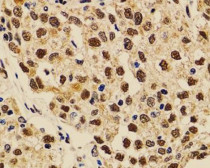
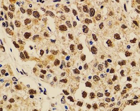
ARG56882 anti-TP53BP1 antibody IHC-P image
Immunohistochemistry: Paraffin-embedded Human lung cancer tissue stained with ARG56882 anti-TP53BP1 antibody at 1:100 dilution.
-

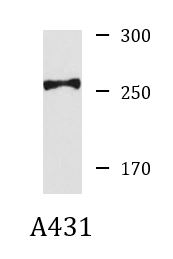
ARG56882 anti-TP53BP1 antibody WB image
Western blot: 25 ug of A431 cell lysate stained with ARG56882 anti-TP53BP1 antibody at 1:1000 dilution.
Customer's Feedback
Good
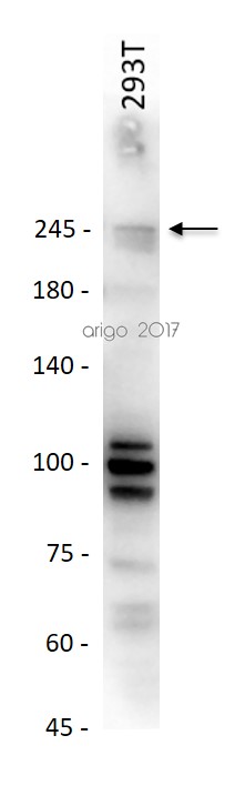
Review for anti-TP53BP1 antibody
Application:WB
Sample:293T
Sample Loading Amount:20 µg
Primary Antibody Dilution Factor:1:500
Primary Antibody Incubation Time:Overnight
Primary Antibody Incubation Temperature:4 ºC


