ARG42491
anti-TP53BP1 antibody
anti-TP53BP1 antibody for Flow cytometry,ICC/IF,IHC-Formalin-fixed paraffin-embedded sections,Western blot and Human,Mouse,Rat
Overview
| Product Description | Rabbit Polyclonal antibody recognizes TP53BP1 |
|---|---|
| Tested Reactivity | Hu, Ms, Rat |
| Tested Application | FACS, ICC/IF, IHC-P, WB |
| Host | Rabbit |
| Clonality | Polyclonal |
| Isotype | IgG |
| Target Name | TP53BP1 |
| Antigen Species | Human |
| Immunogen | Synthetic peptide of Human TP53BP1. |
| Conjugation | Un-conjugated |
| Alternate Names | p53BP1; 53BP1; p202; p53-binding protein 1; Tumor suppressor p53-binding protein 1 |
Application Instructions
| Application Suggestion |
|
||||||||||
|---|---|---|---|---|---|---|---|---|---|---|---|
| Application Note | * The dilutions indicate recommended starting dilutions and the optimal dilutions or concentrations should be determined by the scientist. | ||||||||||
| Positive Control | HeLa |
Properties
| Form | Liquid |
|---|---|
| Purification | Affinity purified. |
| Buffer | PBS (pH 7.4), 150 mM NaCl, 0.02% Sodium azide and 50% Glycerol. |
| Preservative | 0.02% Sodium azide |
| Stabilizer | 50% Glycerol |
| Storage Instruction | For continuous use, store undiluted antibody at 2-8°C for up to a week. For long-term storage, aliquot and store at -20°C. Storage in frost free freezers is not recommended. Avoid repeated freeze/thaw cycles. Suggest spin the vial prior to opening. The antibody solution should be gently mixed before use. |
| Note | For laboratory research only, not for drug, diagnostic or other use. |
Bioinformation
| Database Links |
Swiss-port # Q12888 Human Tumor suppressor p53-binding protein 1 |
|---|---|
| Gene Symbol | TP53BP1 |
| Gene Full Name | tumor protein p53 binding protein 1 |
| Background | This gene encodes a protein that functions in the DNA double-strand break repair pathway choice, promoting non-homologous end joining (NHEJ) pathways, and limiting homologous recombination. This protein plays multiple roles in the DNA damage response, including promoting checkpoint signaling following DNA damage, acting as a scaffold for recruitment of DNA damage response proteins to damaged chromatin, and promoting NHEJ pathways by limiting end resection following a double-strand break. These roles are also important during V(D)J recombination, class switch recombination and at unprotected telomeres. Alternative splicing results in multiple transcript variants encoding different isoforms. [provided by RefSeq, Aug 2017] |
| Function | Double-strand break (DSB) repair protein involved in response to DNA damage, telomere dynamics and class-switch recombination (CSR) during antibody genesis (PubMed:12364621, PubMed:22553214, PubMed:23333306, PubMed:17190600, PubMed:21144835, PubMed:28241136). Plays a key role in the repair of double-strand DNA breaks (DSBs) in response to DNA damage by promoting non-homologous end joining (NHEJ)-mediated repair of DSBs and specifically counteracting the function of the homologous recombination (HR) repair protein BRCA1 (PubMed:22553214, PubMed:23727112, PubMed:23333306). In response to DSBs, phosphorylation by ATM promotes interaction with RIF1 and dissociation from NUDT16L1/TIRR, leading to recruitment to DSBs sites (PubMed:28241136). Recruited to DSBs sites by recognizing and binding histone H2A monoubiquitinated at 'Lys-15' (H2AK15Ub) and histone H4 dimethylated at 'Lys-20' (H4K20me2), two histone marks that are present at DSBs sites (PubMed:23760478, PubMed:28241136, PubMed:17190600). Required for immunoglobulin class-switch recombination (CSR) during antibody genesis, a process that involves the generation of DNA DSBs (PubMed:23345425). Participates to the repair and the orientation of the broken DNA ends during CSR (By similarity). In contrast, it is not required for classic NHEJ and V(D)J recombination (By similarity). Promotes NHEJ of dysfunctional telomeres via interaction with PAXIP1 (PubMed:23727112). [UniProt] |
| Cellular Localization | Nucleus. Chromosome. Chromosome, centromere, kinetochore. Note=Localizes to the nucleus in absence of DNA damage. Following DNA damage, recruited to sites of DNA damage, such as double stand breaks (DSBs): recognizes and binds histone H2A monoubiquitinated at 'Lys-15' (H2AK15Ub) and histone H4 dimethylated at 'Lys-20' (H4K20me2), two histone marks that are present at DSBs sites. Associated with kinetochores during mitosis. [UniProt] |
| Highlight | Related products: TP53BP1 antibodies; Anti-Rabbit IgG secondary antibodies; Related news: Senescence Marker Antibody Panel is launched |
| Calculated MW | 214 kDa |
| PTM | Asymmetrically dimethylated on Arg residues by PRMT1. Methylation is required for DNA binding. Phosphorylated at basal level in the absence of DNA damage (PubMed:11042216, PubMed:11331310). Phosphorylated by ATM in response to DNA damage: phosphorylation at different sites promotes interaction with different set of proteins: phosphorylation at the N-terminus by ATM (residues from 6-178) promotes interaction with PAXIP1 and non-homologous end joining (NHEJ) of dysfunctional telomeres (PubMed:23727112). Phosphorylation by ATM at residues that are located more C-terminus (residues 300-650) leads to promote interaction with RIF1 (PubMed:23727112, PubMed:23333306, PubMed:28241136). Interaction with RIF1 leads to disrupt interaction with NUDT16L1/TIRR (PubMed:28241136). Phosphorylation at Thr-1609 and Ser-1618 in the UDR motif blocks interaction with H2AK15ub (PubMed:24703952). Dephosphorylated by PPP4C (PubMed:24703952). Hyperphosphorylation during mitosis correlates with its exclusion from chromatin and DNA lesions. Hyperphosphorylated in an ATR-dependent manner in response to DNA damage induced by UV irradiation (PubMed:17553757, PubMed:21144835). Dephosphorylated by PPP5C (PubMed:19176521). [UniProt] |
Images (3) Click the Picture to Zoom In
-

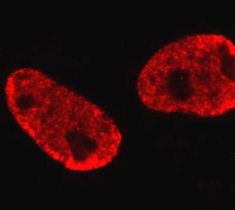
ARG42491 anti-TP53BP1 antibody ICC/IF image
Immunofluorescence: HepG2 cells stained with ARG42491 anti-TP53BP1 antibody.
-
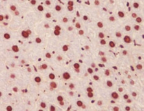
ARG42491 anti-TP53BP1 antibody IHC-P image
Immunohistochemistry: Paraffin-embedded Mouse liver tissue stained with ARG42491 anti-TP53BP1 antibody.
-

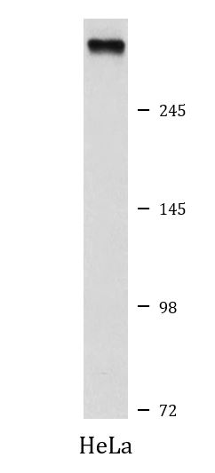
ARG42491 anti-TP53BP1 antibody WB image
Western blot: HeLa cell lysate stained with ARG42491 anti-TP53BP1 antibody.