ARG23741
anti-STAT1 phospho (Tyr701) antibody [SM1351]
anti-STAT1 phospho (Tyr701) antibody [SM1351] for Western blot and Human
Overview
| Product Description | Mouse Monoclonal antibody [SM1351] recognizes STAT1 phospho (Tyr701) |
|---|---|
| Tested Reactivity | Hu |
| Predict Reactivity | Ms, Rat |
| Tested Application | WB |
| Specificity | The antibody detects 84 and 91 kDa stat1 variants on SDS-PAGE immunoblots of human A431 treated with EGF, as well as Jurkat and A431 cells treated with pervanadate. The antibody does not detect these variants in control cells. |
| Host | Mouse |
| Clonality | Monoclonal |
| Clone | SM1351 |
| Isotype | IgG1 |
| Target Name | STAT1 |
| Antigen Species | Human |
| Immunogen | KLH-conjugated phospho-specific peptide around Tyr701 of Human STAT1. This peptide sequence has high homology to the conserved tyrosine site in Rat and Mouse STAT1. |
| Conjugation | Un-conjugated |
| Alternate Names | ISGF-3; Signal transducer and activator of transcription 1-alpha/beta; Transcription factor ISGF-3 components p91/p84; CANDF7; IMD31A; IMD31B; IMD31C; STAT91 |
Application Instructions
| Application Suggestion |
|
||||
|---|---|---|---|---|---|
| Application Note | WB: Antibody is suggested to be diluted in 5% skimmed milk/Tris buffer with 0.04% Tween20 and incubated for 1 hour at room temperature. * The dilutions indicate recommended starting dilutions and the optimal dilutions or concentrations should be determined by the scientist. |
Properties
| Form | Liquid |
|---|---|
| Purification | Purification with Protein A. |
| Buffer | PBS, 0.05% Sodium azide, 50% Glycerol and 1 mg/ml BSA. |
| Preservative | 0.05% Sodium azide |
| Stabilizer | 50% Glycerol and 1 mg/ml BSA |
| Storage Instruction | For continuous use, store undiluted antibody at 2-8°C for up to a week. For long-term storage, aliquot and store at -20°C. Storage in frost free freezers is not recommended. Avoid repeated freeze/thaw cycles. Suggest spin the vial prior to opening. The antibody solution should be gently mixed before use. |
| Note | For laboratory research only, not for drug, diagnostic or other use. |
Bioinformation
| Database Links |
Swiss-port # P42224 Human Signal transducer and activator of transcription 1-alpha/beta |
|---|---|
| Gene Symbol | STAT1 |
| Gene Full Name | signal transducer and activator of transcription 1, 91kDa |
| Background | The protein encoded by this gene is a member of the STAT protein family. In response to cytokines and growth factors, STAT family members are phosphorylated by the receptor associated kinases, and then form homo- or heterodimers that translocate to the cell nucleus where they act as transcription activators. This protein can be activated by various ligands including interferon-alpha, interferon-gamma, EGF, PDGF and IL6. This protein mediates the expression of a variety of genes, which is thought to be important for cell viability in response to different cell stimuli and pathogens. Two alternatively spliced transcript variants encoding distinct isoforms have been described. [provided by RefSeq, Jul 2008] |
| Function | Signal transducer and transcription activator that mediates cellular responses to interferons (IFNs), cytokine KITLG/SCF and other cytokines and other growth factors. Following type I IFN (IFN-alpha and IFN-beta) binding to cell surface receptors, signaling via protein kinases leads to activation of Jak kinases (TYK2 and JAK1) and to tyrosine phosphorylation of STAT1 and STAT2. The phosphorylated STATs dimerize and associate with ISGF3G/IRF-9 to form a complex termed ISGF3 transcription factor, that enters the nucleus. ISGF3 binds to the IFN stimulated response element (ISRE) to activate the transcription of IFN-stimulated genes (ISG), which drive the cell in an antiviral state. In response to type II IFN (IFN-gamma), STAT1 is tyrosine- and serine-phosphorylated. It then forms a homodimer termed IFN-gamma-activated factor (GAF), migrates into the nucleus and binds to the IFN gamma activated sequence (GAS) to drive the expression of the target genes, inducing a cellular antiviral state. Becomes activated in response to KITLG/SCF and KIT signaling. May mediate cellular responses to activated FGFR1, FGFR2, FGFR3 and FGFR4. [UniProt] |
| Highlight | Related products: STAT1 antibodies; STAT1 Duos / Panels; Anti-Mouse IgG secondary antibodies; Related news: Exploring Antiviral Immune Response circNDUFB2, a circular RNA (circRNA), activates anti-tumor immunity |
| Calculated MW | 87 kDa |
| PTM | Phosphorylated on tyrosine and serine residues in response to a variety of cytokines/growth hormones including IFN-alpha, IFN-gamma, PDGF and EGF. Activated KIT promotes phosphorylation on tyrosine residues and subsequent translocation to the nucleus. Upon EGF stimulation, phosphorylation on Tyr-701 (lacking in beta form) by JAK1, JAK2 or TYK2 promotes dimerization and subsequent translocation to the nucleus. Growth hormone (GH) activates STAT1 signaling only via JAK2. Tyrosine phosphorylated in response to constitutively activated FGFR1, FGFR2, FGFR3 and FGFR4. Phosphorylation on Ser-727 by several kinases including MAPK14, ERK1/2 and CAMKII on IFN-gamma stimulation, regulates STAT1 transcriptional activity. Phosphorylation on Ser-727 promotes sumoylation though increasing interaction with PIAS. Phosphorylation on Ser-727 by PRKCD induces apoptosis in response to DNA-damaging agents. Phosphorylated on tyrosine residues when PTK2/FAK1 is activated; most likely this is catalyzed by a SRC family kinase. Dephosphorylation on tyrosine residues by PTPN2 negatively regulates interferon-mediated signaling. Upon viral infection or IFN induction, phosphorylation on Ser-708 occurs much later than phosphorylation on Tyr-701 and is required for the binding of ISGF3 on the ISREs of a subset of IFN-stimulated genes IKBKE-dependent. Phosphorylation at Tyr-701 and Ser-708 are mutually exclusive, phosphorylation at Ser-708 requires previous dephosphorylation of Tyr-701. Sumoylated with SUMO1, SUMO2 and SUMO3. Sumoylation is enhanced by IFN-gamma-induced phosphorylation on Ser-727, and by interaction with PIAS proteins. Enhances the transactivation activity. ISGylated. [UniProt] |
Images (1) Click the Picture to Zoom In
-
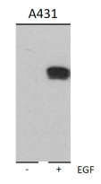
ARG23741 anti-STAT1 phospho (Tyr701) antibody [SM1351] WB image
Western blot: A431 cells untreated (left) or treated with 100 nM EGF for 60 min (right). The blots were stained with ARG23741 anti-STAT1 phospho (Tyr701) antibody [SM1351].


