ARG43095
anti-SRCIN1 / p140Cap antibody
anti-SRCIN1 / p140Cap antibody for Flow cytometry,IHC-Formalin-fixed paraffin-embedded sections,Western blot and Human,Mouse,Rat
Overview
| Product Description | Rabbit Polyclonal antibody recognizes SRCIN1 / p140Cap |
|---|---|
| Tested Reactivity | Hu, Ms, Rat |
| Tested Application | FACS, IHC-P, WB |
| Host | Rabbit |
| Clonality | Polyclonal |
| Isotype | IgG |
| Target Name | SRCIN1 / p140Cap |
| Antigen Species | Human |
| Immunogen | Recombinant protein corresponding to E189-E287 of Human SRCIN1 / p140Cap. |
| Conjugation | Un-conjugated |
| Alternate Names | p130Cas-associated protein; SRC kinase signaling inhibitor 1; SNAP-25-interacting protein; p140Cap; SNIP; P140 |
Application Instructions
| Application Suggestion |
|
||||||||
|---|---|---|---|---|---|---|---|---|---|
| Application Note | IHC-P: Antigen Retrieval: Heat mediation was performed in Citrate buffer (pH 6.0) for 20 min. * The dilutions indicate recommended starting dilutions and the optimal dilutions or concentrations should be determined by the scientist. |
||||||||
| Observed Size | ~ 140 kDa |
Properties
| Form | Liquid |
|---|---|
| Purification | Affinity purification with immunogen. |
| Buffer | 0.2% Na2HPO4, 0.9% NaCl, 0.05% Sodium azide and 4% Trehalose. |
| Preservative | 0.05% Sodium azide |
| Stabilizer | 4% Trehalose |
| Concentration | 0.5 mg/ml |
| Storage Instruction | For continuous use, store undiluted antibody at 2-8°C for up to a week. For long-term storage, aliquot and store at -20°C or below. Storage in frost free freezers is not recommended. Avoid repeated freeze/thaw cycles. Suggest spin the vial prior to opening. The antibody solution should be gently mixed before use. |
| Note | For laboratory research only, not for drug, diagnostic or other use. |
Bioinformation
| Database Links | |
|---|---|
| Gene Symbol | SRCIN1 |
| Gene Full Name | SRC kinase signaling inhibitor 1 |
| Function | Acts as a negative regulator of SRC by activating CSK which inhibits SRC activity and downstream signaling, leading to impaired cell spreading and migration. Regulates dendritic spine morphology. Involved in calcium-dependent exocytosis. May play a role in neurotransmitter release or synapse maintenance. [UniProt] |
| Cellular Localization | Cytoplasm, cytoskeleton. Cell projection, axon, dendrite. Cell junction, synapse, postsynaptic cell membrane, postsynaptic density. Note=Localized to the perinuclear region, lamellopodia, cortical actin and actin stress fibers but not to focal adhesions. Strongly expressed in axons and dendrites of the CA1 and CA3 hippocampal regions and of the dentate gyrus. Detected in both presynapses and postsynapses and in postsynaptic density fractions. [UniProt] |
| Calculated MW | 127 kDa |
| PTM | Tyrosine-phosphorylated in response to EGF and to cell adhesion to integrin ligands. [UniProt] |
Images (11) Click the Picture to Zoom In
-
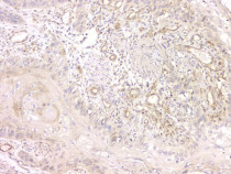
ARG43095 anti-SRCIN1 / p140Cap antibody IHC-P image
Immunohistochemistry: Paraffin-embedded Human appendicitis tissue. Antigen Retrieval: Heat mediation was performed in Citrate buffer (pH 6.0) for 20 min. The tissue section was blocked with 10% goat serum. The tissue section was then stained with ARG43095 anti-SRCIN1 / p140Cap antibody at 1 µg/ml dilution, overnight at 4°C.
-

ARG43095 anti-SRCIN1 / p140Cap antibody WB image
Western blot: 50 µg of sample under reducing conditions. T-47D, MDA-MB-453, Rat brain and Mouse brain lysates stained with ARG43095 anti-SRCIN1 / p140Cap antibody at 0.5 µg/ml dilution, overnight at 4°C.
-
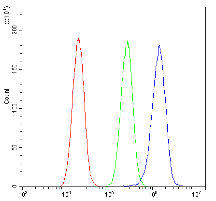
ARG43095 anti-SRCIN1 / p140Cap antibody FACS image
Flow Cytometry: U2OS cells were blocked with 10% normal goat serum and then stained with ARG43095 anti-SRCIN1 / p140Cap antibody (blue) at 1 µg/10^6 cells for 30 min at 20°C, followed by incubation with DyLight®488 labelled secondary antibody. Isotype control antibody (green) was rabbit IgG (1 µg/10^6 cells) used under the same conditions. Unlabelled sample (red) was also used as a control.
-
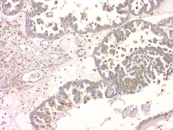
ARG43095 anti-SRCIN1 / p140Cap antibody IHC-P image
Immunohistochemistry: Paraffin-embedded Human ovary cancer tissue. Antigen Retrieval: Heat mediation was performed in Citrate buffer (pH 6.0) for 20 min. The tissue section was blocked with 10% goat serum. The tissue section was then stained with ARG43095 anti-SRCIN1 / p140Cap antibody at 1 µg/ml dilution, overnight at 4°C.
-
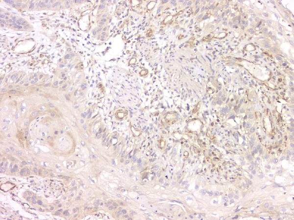
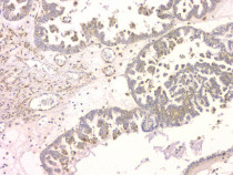
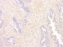
ARG43095 anti-SRCIN1 / p140Cap antibody IHC-P image
Immunohistochemistry: Paraffin-embedded Human oesophagus squama cancer tissue. Antigen Retrieval: Heat mediation was performed in Citrate buffer (pH 6.0) for 20 min. The tissue section was blocked with 10% goat serum. The tissue section was then stained with ARG43095 anti-SRCIN1 / p140Cap antibody at 1 µg/ml dilution, overnight at 4°C.
-
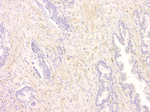
ARG43095 anti-SRCIN1 / p140Cap antibody IHC-P image
Immunohistochemistry: Paraffin-embedded Human ovary cancer tissue. Antigen Retrieval: Heat mediation was performed in Citrate buffer (pH 6.0) for 20 min. The tissue section was blocked with 10% goat serum. The tissue section was then stained with ARG43095 anti-SRCIN1 / p140Cap antibody at 1 µg/ml dilution, overnight at 4°C.
-
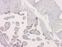
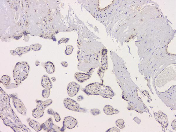
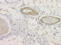
ARG43095 anti-SRCIN1 / p140Cap antibody IHC-P image
Immunohistochemistry: Paraffin-embedded Human placenta tissue. Antigen Retrieval: Heat mediation was performed in Citrate buffer (pH 6.0) for 20 min. The tissue section was blocked with 10% goat serum. The tissue section was then stained with ARG43095 anti-SRCIN1 / p140Cap antibody at 1 µg/ml dilution, overnight at 4°C.
-
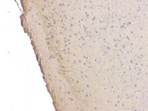
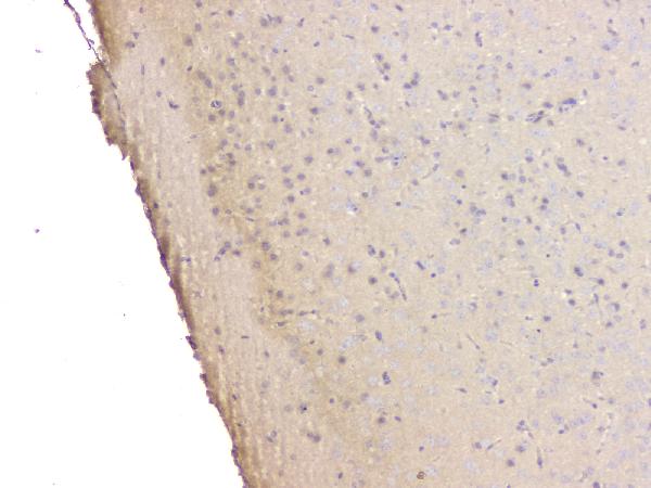
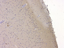
ARG43095 anti-SRCIN1 / p140Cap antibody IHC-P image
Immunohistochemistry: Paraffin-embedded Mouse brain tissue. Antigen Retrieval: Heat mediation was performed in Citrate buffer (pH 6.0) for 20 min. The tissue section was blocked with 10% goat serum. The tissue section was then stained with ARG43095 anti-SRCIN1 / p140Cap antibody at 1 µg/ml dilution, overnight at 4°C.
-
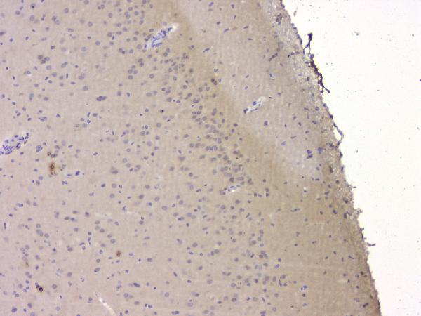
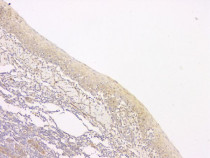
ARG43095 anti-SRCIN1 / p140Cap antibody IHC-P image
Immunohistochemistry: Paraffin-embedded Rat brain tissue. Antigen Retrieval: Heat mediation was performed in Citrate buffer (pH 6.0) for 20 min. The tissue section was blocked with 10% goat serum. The tissue section was then stained with ARG43095 anti-SRCIN1 / p140Cap antibody at 1 µg/ml dilution, overnight at 4°C.
-
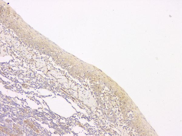
ARG43095 anti-SRCIN1 / p140Cap antibody IHC-P image
Immunohistochemistry: Paraffin-embedded Human tonsil tissue. Antigen Retrieval: Heat mediation was performed in Citrate buffer (pH 6.0) for 20 min. The tissue section was blocked with 10% goat serum. The tissue section was then stained with ARG43095 anti-SRCIN1 / p140Cap antibody at 1 µg/ml dilution, overnight at 4°C.
-

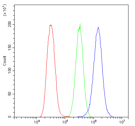
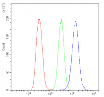
ARG43095 anti-SRCIN1 / p140Cap antibody FACS image
Flow Cytometry: A549 cells were blocked with 10% normal goat serum and then stained with ARG43095 anti-SRCIN1 / p140Cap antibody (blue) at 1 µg/10^6 cells for 30 min at 20°C, followed by incubation with DyLight®488 labelled secondary antibody. Isotype control antibody (green) was rabbit IgG (1 µg/10^6 cells) used under the same conditions. Unlabelled sample (red) was also used as a control.