ARG66123
anti-SF20 antibody (Biotin)
anti-SF20 antibody (Biotin) for ELISA,Western blot and Mouse
Overview
| Product Description | Biotin-conjugated Rabbit Polyclonal antibody recognizes SF20 |
|---|---|
| Tested Reactivity | Ms |
| Tested Application | ELISA, WB |
| Host | Rabbit |
| Clonality | Polyclonal |
| Isotype | IgG |
| Target Name | SF20 |
| Antigen Species | Mouse |
| Immunogen | E. coli derived recombinant Mouse SF20. (MVSEPTTVPF DVRPGGVVHS FSQDVGPGNK FTCTFTYASQ GGTNEQWQMS LGTSEDSQHF TCTIWRPQGK SYLYFTQFKA ELRGAEIEYA MAYSKAAFER ESDVPLKSEE FEVTKTAVSH RPGAFKAELS KLVIVAKAAR SEL) |
| Conjugation | Biotin |
| Alternate Names | EUROIMAGE1875335; IL27w; MYDGF; Interleukin-25; R33729_1; Myeloid-derived growth factor; IL-25; Stromal cell-derived growth factor SF20; IL25; IL27; SF20; C19orf10 |
Application Instructions
| Application Suggestion |
|
||||||
|---|---|---|---|---|---|---|---|
| Application Note | * The dilutions indicate recommended starting dilutions and the optimal dilutions or concentrations should be determined by the scientist. |
Properties
| Form | Liquid |
|---|---|
| Purification | Purified by affinity chromatography. |
| Buffer | PBS (pH 7.2) |
| Concentration | 1 mg/ml |
| Storage Instruction | Aliquot and store in the dark at 2-8°C. Keep protected from prolonged exposure to light. Avoid repeated freeze/thaw cycles. Suggest spin the vial prior to opening. The antibody solution should be gently mixed before use. |
| Note | For laboratory research only, not for drug, diagnostic or other use. |
Bioinformation
| Database Links | |
|---|---|
| Gene Symbol | Mydgf |
| Gene Full Name | myeloid derived growth factor |
| Background | The protein encoded by this gene was previously thought to support proliferation of lymphoid cells and was considered an interleukin. However, this activity has not been reproducible and the function of this protein is currently unknown. [provided by RefSeq, Jul 2008] |
| Function | Bone marrow-derived monocyte and paracrine-acting protein that promotes cardiac myocyte survival and adaptive angiogenesis for cardiac protection and/or repair after myocardial infarction (MI). Stimulates endothelial cell proliferation through a MAPK1/3-, STAT3- and CCND1-mediated signaling pathway. Inhibits cardiac myocyte apoptosis in a PI3K/AKT-dependent signaling pathway (By similarity). Involved in endothelial cell proliferation and angiogenesis. [UniProt] |
| Calculated MW | 19 kDa |
Images (4) Click the Picture to Zoom In
-

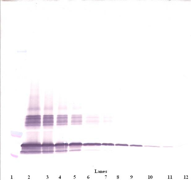
ARG66123 anti-SF20 antibody (Biotin) WB image
Western blot: 250 - 0.24 ng of Mouse SF-20 stained with ARG66123 anti-SF20 antibody (Biotin), under non-reducing conditions.
-
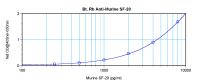
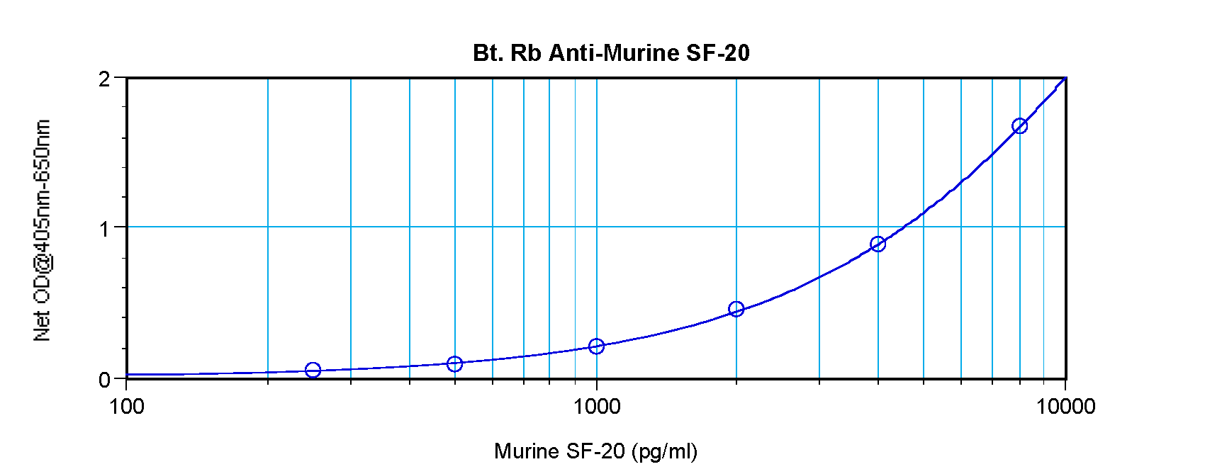
ARG66123 anti-SF20 antibody (Biotin) standard curve image
Direct ELISA: ARG66123 anti-SF20 antibody (Biotin) at 0.25 - 1.0 µg/ml results of a typical standard run with optical density reading at 405 - 650 nm.
-
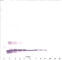
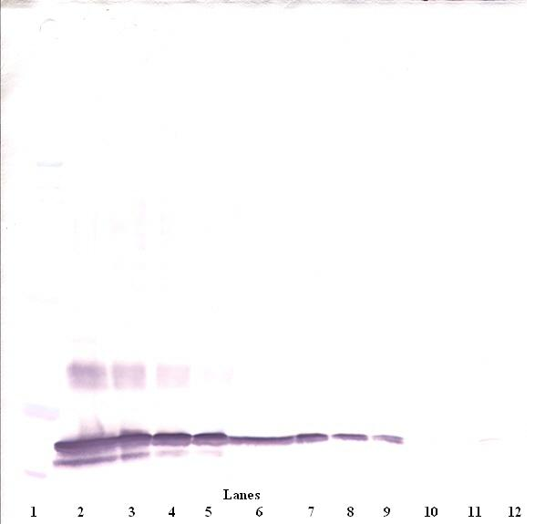
ARG66123 anti-SF20 antibody (Biotin) WB image
Western blot: 250 - 0.24 ng of Mouse SF-20 stained with ARG66123 anti-SF20 antibody (Biotin), under reducing conditions.
-

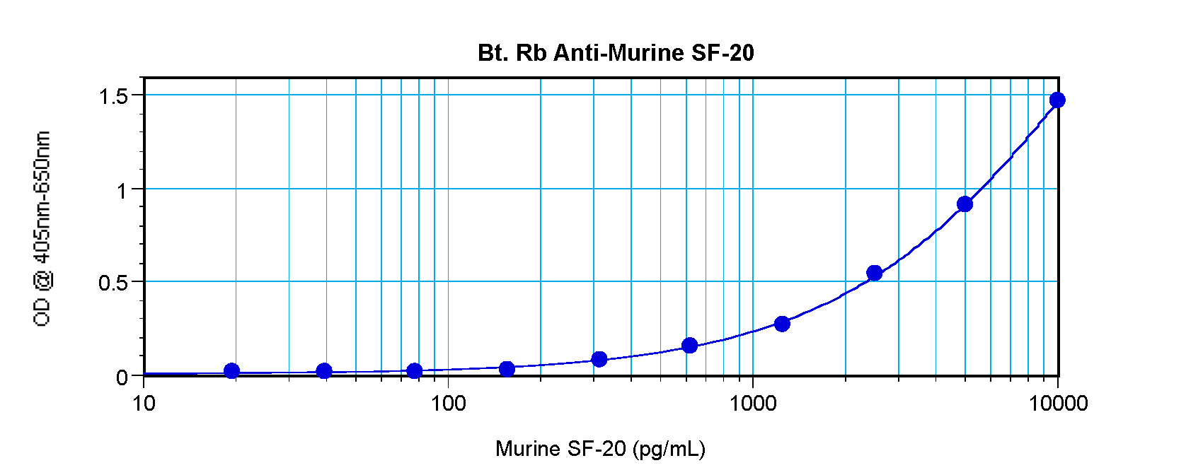
ARG66123 anti-SF20 antibody (Biotin) standard curve image
Sandwich ELISA: ARG66123 anti-SF20 antibody (Biotin) as a detection antibody at 0.25 - 1.0 µg/ml combined with ARG66122 anti-SF20 antibody as a capture antibody. Results of a typical standard run with optical density reading at 405 - 650 nm.