ARG58721
anti-RIPK3 / RIP3 antibody
anti-RIPK3 / RIP3 antibody for ICC/IF,IHC-Formalin-fixed paraffin-embedded sections,Western blot and Human,Mouse,Rat
Overview
| Product Description | Rabbit Polyclonal antibody recognizes RIPK3 / RIP3 |
|---|---|
| Tested Reactivity | Hu, Ms, Rat |
| Tested Application | ICC/IF, IHC-P, WB |
| Host | Rabbit |
| Clonality | Polyclonal |
| Isotype | IgG |
| Target Name | RIPK3 / RIP3 |
| Antigen Species | Human |
| Immunogen | Recombinant fusion protein corresponding to aa. 1-120 of Human RIPK3 / RIP3 (NP_006862.2). |
| Conjugation | Un-conjugated |
| Alternate Names | Receptor-interacting serine/threonine-protein kinase 3; Receptor-interacting protein 3; RIP-3; RIP3; RIP-like protein kinase 3; EC 2.7.11.1 |
Application Instructions
| Application Suggestion |
|
||||||||
|---|---|---|---|---|---|---|---|---|---|
| Application Note | * The dilutions indicate recommended starting dilutions and the optimal dilutions or concentrations should be determined by the scientist. | ||||||||
| Positive Control | Rat lung | ||||||||
| Observed Size | 45-70 kDa |
Properties
| Form | Liquid |
|---|---|
| Purification | Affinity purified. |
| Buffer | PBS (pH 7.3), 0.02% Sodium azide and 50% Glycerol. |
| Preservative | 0.02% Sodium azide |
| Stabilizer | 50% Glycerol |
| Storage Instruction | For continuous use, store undiluted antibody at 2-8°C for up to a week. For long-term storage, aliquot and store at -20°C. Storage in frost free freezers is not recommended. Avoid repeated freeze/thaw cycles. Suggest spin the vial prior to opening. The antibody solution should be gently mixed before use. |
| Note | For laboratory research only, not for drug, diagnostic or other use. |
Bioinformation
| Database Links | |
|---|---|
| Gene Symbol | RIPK3 |
| Gene Full Name | receptor-interacting serine-threonine kinase 3 |
| Background | The product of this gene is a member of the receptor-interacting protein (RIP) family of serine/threonine protein kinases, and contains a C-terminal domain unique from other RIP family members. The encoded protein is predominantly localized to the cytoplasm, and can undergo nucleocytoplasmic shuttling dependent on novel nuclear localization and export signals. It is a component of the tumor necrosis factor (TNF) receptor-I signaling complex, and can induce apoptosis and weakly activate the NF-kappaB transcription factor. [provided by RefSeq, Jul 2008] |
| Function | Essential for necroptosis, a programmed cell death process in response to death-inducing TNF-alpha family members. Upon induction of necrosis, RIPK3 interacts with, and phosphorylates RIPK1 and MLKL to form a necrosis-inducing complex. RIPK3 binds to and enhances the activity of three metabolic enzymes: GLUL, GLUD1, and PYGL. These metabolic enzymes may eventually stimulate the tricarboxylic acid cycle and oxidative phosphorylation, which could result in enhanced ROS production. [UniProt] |
| Cellular Localization | Cytoplasm, cytosol, Cell membrane, Mitochondrion. [UniProt] |
| Highlight | Related products: RIPK3 antibodies; RIPK3 Duos / Panels; Anti-Rabbit IgG secondary antibodies; Related news: RIP1 activation and pathogenesis of NASH Ripoptosome & Necrosome antibody panels are launched |
| Calculated MW | 57 kDa |
| PTM | RIPK1 and RIPK3 undergo reciprocal auto- and trans-phosphorylation. Phosphorylation of Ser-199 plays a role in the necroptotic function of RIPK3. Phosphorylation at Ser-227 is required for binding MLKL. Polyubiquitinated with 'Lys-48' and 'Lys-63'-linked chains by BIRC2/c-IAP1 and BIRC3/c-IAP2, leading to activation of NF-kappa-B. [UniProt] |
Images (2) Click the Picture to Zoom In
-
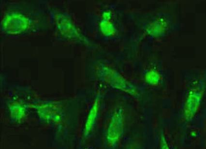
ARG58721 anti-RIPK3 / RIP3 antibody ICC/IF image
Immunofluorescence: U2OS cells stained with ARG58721 anti-RIPK3 / RIP3 antibody at 1:100 dilution.
-

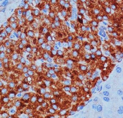
ARG58721 anti-RIPK3 / RIP3 antibody IHC-P image
Immunohistochemistry: Paraffin-embedded Mouse pancreas tissue stained with ARG58721 anti-RIPK3 / RIP3 antibody at 1:100 dilution.