ARG40183
anti-RIPK1 / RIP1 antibody
anti-RIPK1 / RIP1 antibody for ICC/IF,IHC-Formalin-fixed paraffin-embedded sections,Immunoprecipitation,Western blot and Human,Mouse,Rat
Overview
| Product Description | Rabbit Polyclonal antibody recognizes RIPK1 / RIP1 |
|---|---|
| Tested Reactivity | Hu, Ms, Rat |
| Tested Application | ICC/IF, IHC-P, IP, WB |
| Host | Rabbit |
| Clonality | Polyclonal |
| Isotype | IgG |
| Target Name | RIPK1 / RIP1 |
| Antigen Species | Human |
| Immunogen | Recombinant fusion protein corresponding to aa. 170-440 of Human RIPK1 (NP_003795.2). |
| Conjugation | Un-conjugated |
| Alternate Names | Receptor-interacting protein 1; RIP-1; Receptor-interacting serine/threonine-protein kinase 1; RIP; Cell death protein RIP; RIP1; EC 2.7.11.1; Serine/threonine-protein kinase RIP |
Application Instructions
| Application Suggestion |
|
||||||||||
|---|---|---|---|---|---|---|---|---|---|---|---|
| Application Note | * The dilutions indicate recommended starting dilutions and the optimal dilutions or concentrations should be determined by the scientist. | ||||||||||
| Positive Control | C6, Mouse liver and HT-1080 | ||||||||||
| Observed Size | 76 kDa |
Properties
| Form | Liquid |
|---|---|
| Purification | Affinity purified. |
| Buffer | PBS (pH 7.3), 0.02% Sodium azide and 50% Glycerol. |
| Preservative | 0.02% Sodium azide |
| Stabilizer | 50% Glycerol |
| Storage Instruction | For continuous use, store undiluted antibody at 2-8°C for up to a week. For long-term storage, aliquot and store at -20°C. Storage in frost free freezers is not recommended. Avoid repeated freeze/thaw cycles. Suggest spin the vial prior to opening. The antibody solution should be gently mixed before use. |
| Note | For laboratory research only, not for drug, diagnostic or other use. |
Bioinformation
| Database Links |
Swiss-port # Q13546 Human Receptor-interacting serine/threonine-protein kinase 1 Swiss-port # Q60855 Mouse Receptor-interacting serine/threonine-protein kinase 1 |
|---|---|
| Gene Symbol | RIPK1 |
| Gene Full Name | receptor (TNFRSF)-interacting serine-threonine kinase 1 |
| Background | RIPK1 / RIP1 is a member of the receptor-interacting protein (RIP) family of serine/threonine protein kinases. The encoded protein plays a role in inflammation and cell death in response to tissue damage, pathogen recognition, and as part of developmental regulation. RIPK1/RIPK3 kinase-mediated necrosis is referred to as necroptosis. Genetic disruption of this gene in mice results in death shortly after birth. [provided by RefSeq, Aug 2017] |
| Function | RIPK1 / RIP1: Serine-threonine kinase which is a key regulator of both cell death and cell survival (PubMed:25459879). Exhibits kinase activity-dependent functions that trigger cell death and kinase-independent scaffold functions regulating inflammatory signaling and cell survival (PubMed:11101870, PubMed:25459879). Initiates ripoptocide which describes cell death that is dependent on RIPK1, be it apoptosis or necroptosis (PubMed:31457011). Upon binding of TNF to TNFR1, RIPK1 is recruited to the TNF-R1 signaling complex (TNF-RSC also known as complex I) where it acts as a scaffold protein promoting cell survival, in part, by activating the canonical NF-kB pathway. Specific conditions can however activate RIPK1, and its kinase activity then regulates assembly of two death-inducing complexes, namely complex IIa (RIPK1-FADD-CASP8) and the complex IIb (RIPK1-RIPK3-MLKL) and these complexes respectively drive apoptosis or necroptosis, a regulated form of necrosis (PubMed:19524513, PubMed:19524512, PubMed:29440439, PubMed:30988283). During embryonic development suppresses apoptosis and necroptosis and prevents the interaction of TRADD with FADD thereby limiting aberrant activation of CASP8. Phosphorylates DAB2IP at 'Ser-728' in a TNF- alpha-dependent manner, and thereby activates the MAP3K5-JNK apoptotic cascade (PubMed:17389591). Required for ZBP1-induced NF-kappaB activation and activation of NF-kappaB by DNA damage and IR. [UniProt] |
| Cellular Localization | Cytoplasm, Cell membrane. [UniProt] |
| Highlight | Related products: RIPK1 antibodies; RIPK1 Duos / Panels; Anti-Rabbit IgG secondary antibodies; Related news: RIP1 activation and pathogenesis of NASH Ripoptosome & Necrosome antibody panels are launched |
| Calculated MW | 76 kDa |
| PTM | Proteolytically cleaved by caspase-8 during TNF-induced apoptosis. Cleavage abolishes NF-kappa-B activation and enhances pro-apoptotic signaling through the TRADD-FADD interaction. RIPK1 and RIPK3 undergo reciprocal auto- and trans-phosphorylation. Phosphorylation of Ser-161 by RIPK3 is necessary for the formation of the necroptosis-inducing complex. Ubiquitinated by 'Lys-11'-, 'Lys-48'-, 'Lys-63'- and linear-linked type ubiquitin. Polyubiquitination with 'Lys-63'-linked chains by TRAF2 induces association with the IKK complex. Deubiquitination of 'Lys-63'-linked chains and polyubiquitination with 'Lys-48'-linked chains by TNFAIP3 leads to RIPK1 proteasomal degradation and consequently down-regulates TNF-alpha-induced NFkappa-B signaling. 'Lys-48'-linked polyubiquitination by RFFL or RNF34 also promotes proteasomal degradation and negatively regulates TNF-alpha-induced NFkappa-B signaling. Linear polyubiquitinated; the head-to-tail polyubiquitination is mediated by the LUBAC complex. LPS-mediated activation of NF-kappa-B. Also ubiquitinated with 'Lys-11'-linked chains. Polyubiquitinated with 'Lys-48' and 'Lys-63'-linked chains by BIRC2/c-IAP1 and BIRC3/c-IAP2, leading to activation of NF-kappa-B. [UniProt] |
Images (4) Click the Picture to Zoom In
-
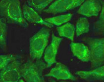
ARG40183 anti-RIPK1 / RIP1 antibody ICC/IF image
Immunofluorescence: U2OS cells stained with ARG40183 anti-RIPK1 / RIP1 antibody at 1:100 dilution.
-
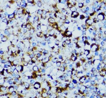
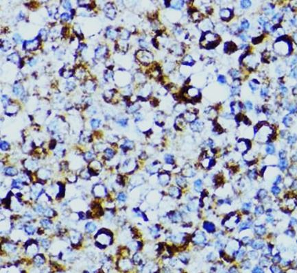
ARG40183 anti-RIPK1 / RIP1 antibody IHC-P image
Immunohistochemistry: Paraffin-embedded Human tonsil tissue stained with ARG40183 anti-RIPK1 / RIP1 antibody at 1:100 dilution.
-
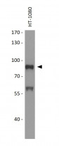
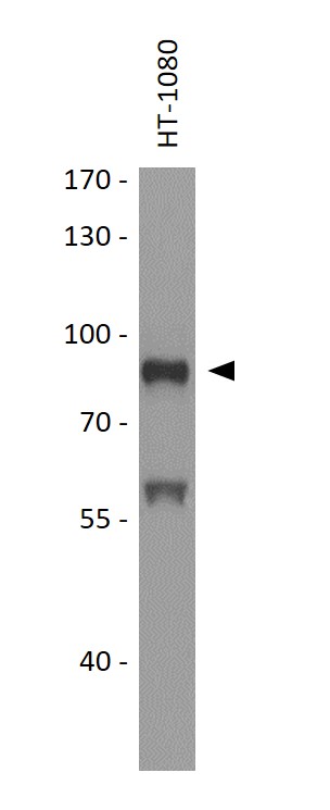
ARG40183 anti-RIPK1 / RIP1 antibody WB image
Western blot: 25 µg of HT-1080 cell lysate stained with ARG40183 anti-RIPK1 / RIP1 antibody at 1:1000 dilution.
-
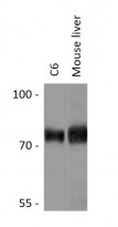
ARG40183 anti-RIPK1 / RIP1 antibody WB image
Western blot: 25 µg of C6 and Mouse liver lysates stained with ARG40183 anti-RIPK1 / RIP1 antibody at 1:1000 dilution.


Specific References
Silencing of TRAF5 enhances necroptosis in hepatocellular carcinoma by inhibiting LTBR-mediated NF-κB signaling
WB / Human


