ARG63724
anti-Plexin D1 antibody
anti-Plexin D1 antibody for Flow cytometry,ICC/IF and Human
Cell Biology and Cellular Response antibody
Overview
| Product Description | Goat Polyclonal antibody recognizes Plexin D1 |
|---|---|
| Tested Reactivity | Hu |
| Predict Reactivity | Cow, Dog, Pig |
| Tested Application | FACS, ICC/IF |
| Host | Goat |
| Clonality | Polyclonal |
| Isotype | IgG |
| Target Name | Plexin D1 |
| Antigen Species | Human |
| Immunogen | C-LAEPKKSHRQSH |
| Conjugation | Un-conjugated |
| Alternate Names | PLEXD1; Plexin-D1 |
Application Instructions
| Application Suggestion |
|
||||||
|---|---|---|---|---|---|---|---|
| Application Note | * The dilutions indicate recommended starting dilutions and the optimal dilutions or concentrations should be determined by the scientist. |
Properties
| Form | Liquid |
|---|---|
| Purification | Purified from goat serum by antigen affinity chromatography. |
| Buffer | Tris saline (pH 7.3), 0.02% Sodium azide and 0.5% BSA. |
| Preservative | 0.02% Sodium azide |
| Stabilizer | 0.5% BSA |
| Concentration | 0.5 mg/ml |
| Storage Instruction | For continuous use, store undiluted antibody at 2-8°C for up to a week. For long-term storage, aliquot and store at -20°C or below. Storage in frost free freezers is not recommended. Avoid repeated freeze/thaw cycles. Suggest spin the vial prior to opening. The antibody solution should be gently mixed before use. |
| Note | For laboratory research only, not for drug, diagnostic or other use. |
Bioinformation
| Database Links | |
|---|---|
| Gene Symbol | PLXND1 |
| Gene Full Name | plexin D1 |
| Function | Cell surface receptor for SEMA4A and for class 3 semaphorins, such as SEMA3A, SEMA3C and SEMA3E. Plays an important role in cell-cell signaling, and in regulating the migration of a wide spectrum of cell types. Regulates the migration of thymocytes in the medulla. Regulates endothelial cell migration. Plays an important role in ensuring the specificity of synapse formation. Required for normal development of the heart and vasculature (By similarity). Mediates anti-angiogenic signaling in response to SEMA3E. [UniProt] |
| Research Area | Cell Biology and Cellular Response antibody |
| Calculated MW | 212 kDa |
Images (2) Click the Picture to Zoom In
-
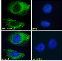
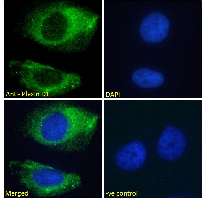
ARG63724 anti-Plexin D1 antibody ICC/IF image
Immunofluorescence: Paraformaldehyde fixed HeLa cells permeabilized with 0.15% Triton. Cells were stained with ARG63724 anti-Plexin D1 antibody (green) at 10 µg/ml dilution for 1 hour. DAPI (blue) for nuclear staining. Negative control: Unimmunized goat IgG (green) at 10 µg/ml dilution.
-
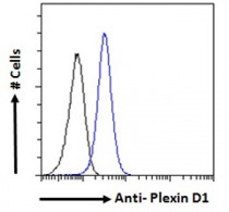
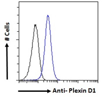
ARG63724 anti-Plexin D1 antibody FACS image
Flow Cytometry: Paraformaldehyde-fixed K562 cells permeabilized with 0.5% Triton. Cells were stained with ARG63724 anti-Plexin D1 antibody (blue line) at 10 µg/ml dilution for 1 hour, followed by incubation with Alexa FluorR 488 labelled secondary antibody. IgG control: Unimmunized goat IgG (black line).