ARG11131
anti-Peripherin antibody [8G2]
anti-Peripherin antibody [8G2] for ICC/IF,IHC-Frozen sections,Western blot and Human,Mouse,Rat,Cow,Pig
Overview
| Product Description | Mouse Monoclonal antibody [8G2] recognizes Peripherin |
|---|---|
| Tested Reactivity | Hu, Ms, Rat, Cow, Pig |
| Tested Application | ICC/IF, IHC-Fr, WB |
| Host | Mouse |
| Clonality | Monoclonal |
| Clone | 8G2 |
| Isotype | IgG1 |
| Target Name | Peripherin |
| Antigen Species | Rat |
| Immunogen | Recombinant Rat Peripherin. |
| Conjugation | Un-conjugated |
| Alternate Names | Peripherin; NEF4; PRPH1; Neurofilament 4 |
Application Instructions
| Application Suggestion |
|
||||||||
|---|---|---|---|---|---|---|---|---|---|
| Application Note | * The dilutions indicate recommended starting dilutions and the optimal dilutions or concentrations should be determined by the scientist. | ||||||||
| Observed Size | ~ 54 kDa |
Properties
| Form | Liquid |
|---|---|
| Purification | Purified |
| Buffer | PBS, 5 mM Sodium azide and 50% Glycerol. |
| Preservative | 5 mM Sodium azide |
| Stabilizer | 50% Glycerol |
| Concentration | 1 mg/ml |
| Storage Instruction | For continuous use, store undiluted antibody at 2-8°C for up to a week. For long-term storage, aliquot and store at -20°C. Storage in frost free freezers is not recommended. Avoid repeated freeze/thaw cycles. Suggest spin the vial prior to opening. The antibody solution should be gently mixed before use. |
| Note | For laboratory research only, not for drug, diagnostic or other use. |
Bioinformation
| Database Links | |
|---|---|
| Gene Symbol | PRPH |
| Gene Full Name | peripherin |
| Background | This gene encodes a cytoskeletal protein found in neurons of the peripheral nervous system. The encoded protein is a type III intermediate filament protein with homology to other cytoskeletal proteins such as desmin, and is a different protein that the peripherin found in photoreceptors. Mutations in this gene have been associated with susceptibility to amyotrophic lateral sclerosis. [provided by RefSeq, Jul 2008] |
| Function | Class-III neuronal intermediate filament protein. [UniProt] |
| Calculated MW | 54 kDa |
Images (3) Click the Picture to Zoom In
-
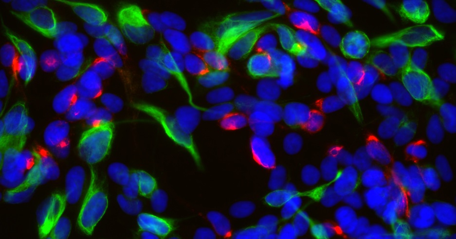
ARG11131 anti-Peripherin antibody [8G2] ICC/IF image
Immunofluorescence: SH-SY5Y cells stained with ARG11131 anti-Peripherin antibody [8G2] (red) at 1:500 dilution, and co-stained with anti-Vimentin antibody (green) at 1:10000 dilution. DAPI (blue) for nuclear staining.
-

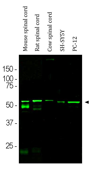
ARG11131 anti-Peripherin antibody [8G2] WB image
Western blot: Mouse spinal cord, Rat spinal cord, Cow spinal cord, SH-SY5Y and PC-12 cell lysates stained with ARG11131 anti-Peripherin antibody [8G2] at 1:500 dilution.
The band at ~ 57 kDa corresponds to the peripherin protein. Bands at 50 and 25 kDa detected in the mouse spinal cord lysate, correspond to the heavy and light chains of mouse IgG.
-
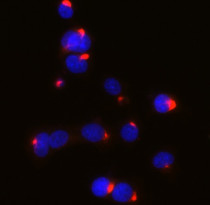
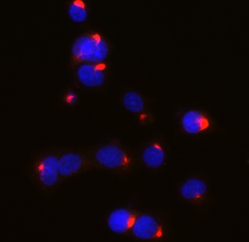
ARG11131 anti-Peripherin antibody [8G2] ICC/IF image
Immunofluorescence: PC-12 cells stained with ARG11131 anti-Peripherin antibody [8G2] (red) at 1:500 dilution, and co-stained with anti-Vimentin antibody (green) at 1:10000 dilution. DAPI (blue) for nuclear staining.