ARG63196
anti-NQO1 antibody
anti-NQO1 antibody for ICC/IF,IHC-Formalin-fixed paraffin-embedded sections,Western blot and Human,Rat,Pig
Metabolism antibody; Signaling Transduction antibody

Overview
| Product Description | Goat Polyclonal antibody recognizes NQO1 |
|---|---|
| Tested Reactivity | Hu, Rat, Pig |
| Predict Reactivity | Dog |
| Tested Application | ICC/IF, IHC-P, WB |
| Specificity | This products is expected to recognize all three reported isoforms (NP_000894.1, NP_001020604.1 and NP_001020605.1). |
| Host | Goat |
| Clonality | Polyclonal |
| Isotype | IgG |
| Target Name | NQO1 |
| Antigen Species | Human |
| Immunogen | C-SIPTDNQIKARK |
| Conjugation | Un-conjugated |
| Alternate Names | NMOR1; EC 1.6.5.2; NAD; DHQU; P; Quinone reductase 1; DT-diaphorase; DTD; QR1; Phylloquinone reductase; NMORI; DIA4; Menadione reductase; Azoreductase |
Application Instructions
| Application Suggestion |
|
||||||||
|---|---|---|---|---|---|---|---|---|---|
| Application Note | WB: Recommend incubate at RT for 1h. IHC-P: Antigen Retrieval: Steam tissue section in Citrate buffer (pH 6.0). * The dilutions indicate recommended starting dilutions and the optimal dilutions or concentrations should be determined by the scientist. |
Properties
| Form | Liquid |
|---|---|
| Purification | Purified from goat serum by antigen affinity chromatography. |
| Buffer | Tris saline (pH 7.3), 0.02% Sodium azide and 0.5% BSA. |
| Preservative | 0.02% Sodium azide |
| Stabilizer | 0.5% BSA |
| Concentration | 0.5 mg/ml |
| Storage Instruction | For continuous use, store undiluted antibody at 2-8°C for up to a week. For long-term storage, aliquot and store at -20°C or below. Storage in frost free freezers is not recommended. Avoid repeated freeze/thaw cycles. Suggest spin the vial prior to opening. The antibody solution should be gently mixed before use. |
| Note | For laboratory research only, not for drug, diagnostic or other use. |
Bioinformation
| Database Links | |
|---|---|
| Background | This gene is a member of the NAD(P)H dehydrogenase (quinone) family and encodes a cytoplasmic 2-electron reductase. This FAD-binding protein forms homodimers and reduces quinones to hydroquinones. This protein's enzymatic activity prevents the one electron reduction of quinones that results in the production of radical species. Mutations in this gene have been associated with tardive dyskinesia (TD), an increased risk of hematotoxicity after exposure to benzene, and susceptibility to various forms of cancer. Altered expression of this protein has been seen in many tumors and is also associated with Alzheimer's disease (AD). Alternate transcriptional splice variants, encoding different isoforms, have been characterized. [provided by RefSeq, Jul 2008] |
| Highlight | Related products: NQO1 antibodies; NQO1 Duos / Panels; Anti-Goat IgG secondary antibodies; Related news: Keap1-Nrf2-ARE antibody panel is launched |
| Research Area | Metabolism antibody; Signaling Transduction antibody |
| Calculated MW | 31 kDa |
Images (6) Click the Picture to Zoom In
-
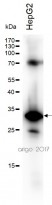
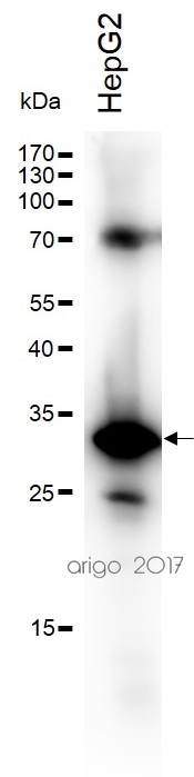
ARG63196 anti-NQO1 antibody WB image
Western blot: 30 µg of HepG2 cell lysate stained with ARG63196 anti-NQO1 antibody at 1:500 dilution.
-
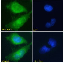
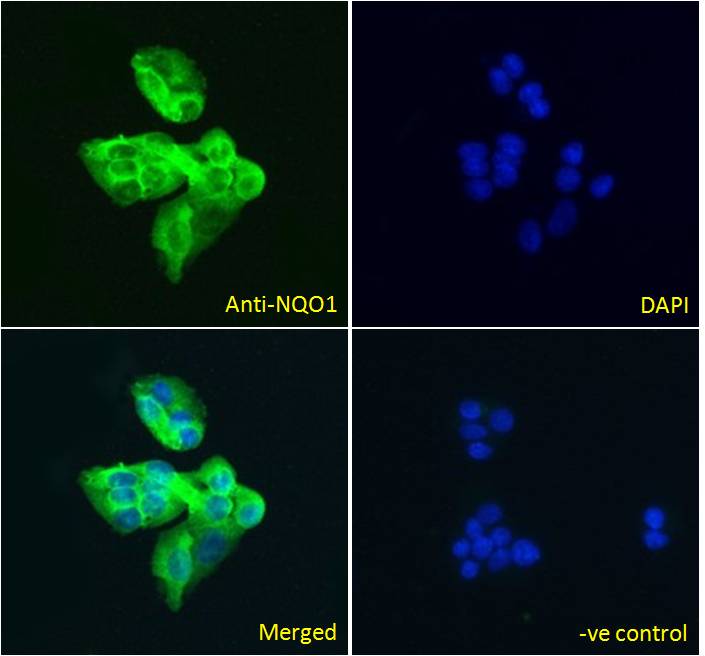
ARG63196 anti-NQO1 antibody ICC/IF image
Immunofluorescence: Paraformaldehyde fixed U251 cells permeabilized with 0.15% Triton. Cells were stained with ARG63196 anti-NQO1 antibody (green) at 10 µg/ml dilution for 1 hour. DAPI (blue) for nuclear staining. Negative control: Unimmunized goat IgG (green) at 10 µg/ml dilution.
-

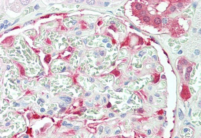
ARG63196 anti-NQO1 antibody IHC-P image
Immunohistochemistry: Paraffin-embedded Human kidney tissue. Antigen Retrieval: Steam tissue section in Citrate buffer (pH 6.0). The tissue section was stained with ARG63196 anti-NQO1 antibody at 5 µg/ml dilution followed by AP-staining.
-

ARG63196 anti-NQO1 antibody WB image
Western blot: 35 µg of U251 cell lysate (in RIPA buffer) stained with ARG63196 anti-NQO1 antibody at 0.1 µg/ml dilution and incubated at RT for 1 hour.
-
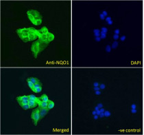
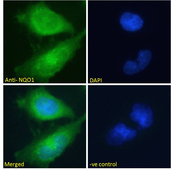
ARG63196 anti-NQO1 antibody ICC/IF image
Immunofluorescence: Paraformaldehyde fixed HepG2 cells permeabilized with 0.15% Triton. Cells were stained with ARG63196 anti-NQO1 antibody (green) at 5 µg/ml dilution for 1 hour. DAPI (blue) for nuclear staining. Negative control: Unimmunized goat IgG (green) at 5 µg/ml dilution.
-
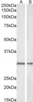
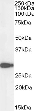
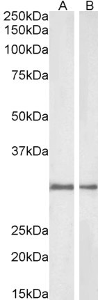
ARG63196 anti-NQO1 antibody WB image
Western blot: 35 µg of Rat (A) and Pig (B) kidney lysates (in RIPA buffer) stained with ARG63196 anti-NQO1 antibody at 1 µg/ml dilution and incubated at RT for 1 hour.
Customer's Feedback
Good
Review for anti-NQO1 antibody
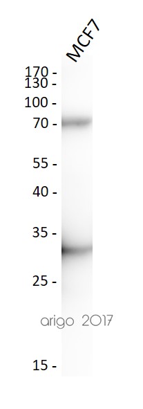
Application:WB
Sample:MCF7
Sample Loading Amount:30 µg
Primary Antibody Dilution Factor:1:500
Primary Antibody Incubation Time:overnight
Primary Antibody Incubation Temperature:4 ºC


