ARG44682
anti-Mitofilin antibody
anti-Mitofilin antibody for IHC-Formalin-fixed paraffin-embedded sections,Immunoprecipitation,Western blot and Human
Overview
| Product Description | Mouse Monoclonal antibody recognizes Mitofilin |
|---|---|
| Tested Reactivity | Hu |
| Tested Application | IHC-P, IP, WB |
| Host | Mouse |
| Clonality | Monoclonal |
| Isotype | IgG1 |
| Target Name | Mitofilin |
| Antigen Species | Human |
| Conjugation | Un-conjugated |
| Alternate Names | Cell proliferation-inducing gene 4/52 protein; P89; MICOS complex subunit MIC60; PIG52; P87/89; HMP; P87; Mitofilin; Mic60; PIG4; p87/89; MINOS2; Mitochondrial inner membrane protein |
Application Instructions
| Application Suggestion |
|
||||||||
|---|---|---|---|---|---|---|---|---|---|
| Application Note | * The dilutions indicate recommended starting dilutions and the optimal dilutions or concentrations should be determined by the scientist. |
Properties
| Form | Liquid |
|---|---|
| Purification | Protein A purification |
| Buffer | PBS with 0.09% sodium azide |
| Storage Instruction | For continuous use, store undiluted antibody at 2-8°C for up to a week. For long-term storage, aliquot and store at -20°C or below. Storage in frost free freezers is not recommended. Avoid repeated freeze/thaw cycles. Suggest spin the vial prior to opening. The antibody solution should be gently mixed before use. |
| Note | For laboratory research only, not for drug, diagnostic or other use. |
Bioinformation
| Database Links | |
|---|---|
| Gene Symbol | IMMT |
| Gene Full Name | inner membrane protein, mitochondrial |
| Function | Component of the MICOS complex, a large protein complex of the mitochondrial inner membrane that plays crucial roles in the maintenance of crista junctions, inner membrane architecture, and formation of contact sites to the outer membrane. Plays an important role in the maintenance of the MICOS complex stability and the mitochondrial cristae morphology. [UniProt] |
| Cellular Localization | Mitochondrion inner membrane; Single-pass membrane protein |
| Calculated MW | 84 kDa |
| PTM | N-glycosylation enhances cell surface expression and lengthens receptor half-life by preventing degradation in the ER. |
Images (3) Click the Picture to Zoom In
-
ARG44682 anti-Mitofilin antibody IHC-P image
Immunohistochemistry: Human Kidney stained with ARG44682 anti-Mitofilin antibody at 5 µg/mL dilution.
-
ARG44682 anti-Mitofilin antibody WB image
Western blot: Raji stained with ARG44682 anti-Mitofilin antibody at 1 µg/mL dilution.
-
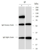
ARG44682 anti-Mitofilin antibody IP image
Immunoprecipitation: MCF7 lysate immunoprecipitated with 2.5 µg of ARG44682 anti-Mitofilin antibody.


