ARG54860
anti-MLKL antibody
anti-MLKL antibody for Western blot and Mouse
Cancer antibody; Signaling Transduction antibody
Overview
| Product Description | Rabbit Polyclonal antibody recognizes MLKL |
|---|---|
| Tested Reactivity | Ms |
| Tested Application | WB |
| Host | Rabbit |
| Clonality | Polyclonal |
| Isotype | IgG |
| Target Name | MLKL |
| Antigen Species | Mouse |
| Immunogen | KLH-conjugated synthetic peptide corresponding to aa. 444-472 (C-terminus) of Mouse MLKL. |
| Conjugation | Un-conjugated |
| Alternate Names | Mixed lineage kinase domain-like protein; hMLKL |
Application Instructions
| Application Suggestion |
|
||||
|---|---|---|---|---|---|
| Application Note | * The dilutions indicate recommended starting dilutions and the optimal dilutions or concentrations should be determined by the scientist. | ||||
| Positive Control | Mouse lung |
Properties
| Form | Liquid |
|---|---|
| Purification | Purification with Protein A and immunogen peptide. |
| Buffer | PBS and 0.09% (W/V) Sodium azide |
| Preservative | 0.09% (W/V) Sodium azide |
| Storage Instruction | For continuous use, store undiluted antibody at 2-8°C for up to a week. For long-term storage, aliquot and store at -20°C or below. Storage in frost free freezers is not recommended. Avoid repeated freeze/thaw cycles. Suggest spin the vial prior to opening. The antibody solution should be gently mixed before use. |
| Note | For laboratory research only, not for drug, diagnostic or other use. |
Bioinformation
| Database Links |
Swiss-port # Q9D2Y4 Mouse Mixed lineage kinase domain-like protein |
|---|---|
| Gene Symbol | Mlkl |
| Gene Full Name | mixed lineage kinase domain-like |
| Background | This gene belongs to the protein kinase superfamily. The encoded protein contains a protein kinase-like domain; however, is thought to be inactive because it lacks several residues required for activity. This protein plays a critical role in tumor necrosis factor (TNF)-induced necroptosis, a programmed cell death process, via interaction with receptor-interacting protein 3 (RIP3), which is a key signaling molecule in necroptosis pathway. Inhibitor studies and knockdown of this gene inhibited TNF-induced necrosis. High levels of this protein and RIP3 are associated with inflammatory bowel disease in children. Alternatively spliced transcript variants have been described for this gene. [provided by RefSeq, Sep 2015] |
| Function | Pseudokinase that plays a key role in TNF-induced necroptosis, a programmed cell death process. Activated following phosphorylation by RIPK3, leading to homotrimerization, localization to the plasma membrane and execution of programmed necrosis characterized by calcium influx and plasma membrane damage. Does not have protein kinase activity. [UniProt] |
| Cellular Localization | Cytoplasm. Cell membrane. Note=Localizes to the cytoplasm and translocates to the plasma membrane on necroptosis induction. |
| Highlight | Related products: MLKL antibodies; MLKL Duos / Panels; Anti-Rabbit IgG secondary antibodies; Related news: RIP1 activation and pathogenesis of NASH Ripoptosome & Necrosome antibody panels are launched |
| Research Area | Cancer antibody; Signaling Transduction antibody |
| Calculated MW | 54 kDa |
| PTM | Phosphorylation by RIPK3 induces a conformational switch that is required for necroptosis. It also induces homotrimerization and localization to the plasma membrane. |
Images (1) Click the Picture to Zoom In
-

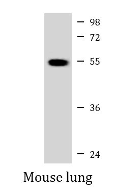
ARG54860 anti-MLKL antibody WB image
Western blot: 35 µg of Mouse lung tissue lysate stained with ARG54860 anti-MLKL antibody at 1:1000 dilution.