ARG66982
anti-Ki-67 antibody [SP6]
anti-Ki-67 antibody [SP6] for ICC/IF,IHC-Formalin-fixed paraffin-embedded sections,Western blot and Human,Mouse,Rat
Microvascular Density Study antibody; Proliferation Marker antibody
Overview
| Product Description | Rabbit Monoclonal antibody [SP6] recognizes Ki-67 |
|---|---|
| Tested Reactivity | Hu, Ms, Rat |
| Tested Application | ICC/IF, IHC-P, WB |
| Host | Rabbit |
| Clonality | Monoclonal |
| Clone | SP6 |
| Isotype | IgG |
| Target Name | Ki-67 |
| Antigen Species | Human |
| Immunogen | Synthetic peptide from C-terminus of human Ki-67 protein. |
| Conjugation | Un-conjugated |
| Alternate Names | Antigen KI-67; MIB-; KIA; MIB-1; PPP1R105 |
Application Instructions
| Application Suggestion |
|
||||||||
|---|---|---|---|---|---|---|---|---|---|
| Application Note | IHC-P: Antigen Retrieval: Boil tissue section in 10mM citrate buffer, pH 6.0 for 10 min followed by cooling at RT for 20 min. Incubation Time: 30 min at RT. * The dilutions indicate recommended starting dilutions and the optimal dilutions or concentrations should be determined by the scientist. |
Properties
| Form | Liquid |
|---|---|
| Purification | Affinity purified. |
| Buffer | 100 mM Tris Glycine (pH 7.0), 0.025% ProClin 300, 1%BSA and 20% Glycerol. |
| Preservative | 0.025% ProClin 300 |
| Stabilizer | 1%BSA and 20% Glycerol |
| Storage Instruction | For continuous use, store undiluted antibody at 2-8°C for up to a week. For long-term storage, aliquot and store at -20°C or below. Storage in frost free freezers is not recommended. Avoid repeated freeze/thaw cycles. Suggest spin the vial prior to opening. The antibody solution should be gently mixed before use. |
| Note | For laboratory research only, not for drug, diagnostic or other use. |
Bioinformation
| Database Links | |
|---|---|
| Gene Symbol | MKI67 |
| Gene Full Name | marker of proliferation Ki-67 |
| Background | Ki-67 is a nuclear protein, which is expressed in the proliferating cells. Ki-67 is preferentially expressed during late G1-, S-, M-, and G2-phases of the cell cycle, while cells in the G0 (quiescent) phase are negative for this protein. |
| Cellular Localization | Nucleus |
| Highlight | Related products: Ki67 antibodies; Ki67 Duos / Panels; Anti-Rabbit IgG secondary antibodies; Related news: Understanding Your Cells: Choose the right markers SM5-1, a promising immunotherapy for Hepatocellular Carcinoma (HCC) Neuronal Development Marker Choose the Best ZIKA Virus Antibodies Fight microcephaly with arigo Tools for studying Exosomes |
| Research Area | Microvascular Density Study antibody; Proliferation Marker antibody |
| Calculated MW | 345 kDa and 395 kDa |
| PTM | Phosphorylated. Hyperphosphorylated in mitosis (PubMed:10502411, PubMed:10653604). Hyperphosphorylated form does not bind DNA. |
Images (3) Click the Picture to Zoom In
-
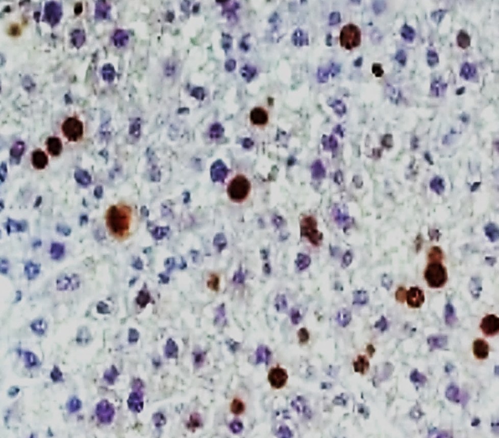
ARG66982 anti-Ki-67 antibody IHC-P image
Immunohistochemistry: Paraffin-embedded Mouse liver tissue stained with ARG66982 anti-Ki-67 antibody at 1:100 dilution.
-
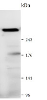
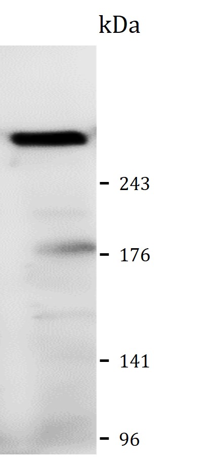
ARG66982 anti-Ki-67 antibody WB image
Western blot: CT26 cell lysate stained with ARG66982 anti-Ki-67 antibody at 1:500 dilution, overnight at 4°C.
-

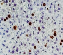
ARG66982 anti-Ki-67 antibody IHC-P image
Immunohistochemistry: Paraffin-embedded Mouse heart tissue stained with ARG66982 anti-Ki-67 antibody at 1:100 dilution.