Understanding Your Cells: Choose the right markers
Understanding Your Cells: Choose the right markers
|
Understanding what’s going on in a cell or tissue type is of particular interest for all scientists who study cell biology. Most cells express a certain set of proteins and markers when they undergo different conditions which lead them to exhibit certain phenotypes. In order to understand what the cells are going through, we need to probe our samples with the right antibodies. What kind of markers or antibodies should I use for the detection of apoptosis instead of autophagy? Under certain circumstances, how would I know if my cells are undergoing proliferation or cell-cycle arrest? Read on, and you’ll find the most suitable markers used to probe for some common signals sent out by cells in response to certain stimulus.
|
||
 |
 |
When the cells are under attack |
 |
||
|
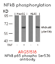
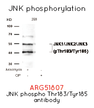
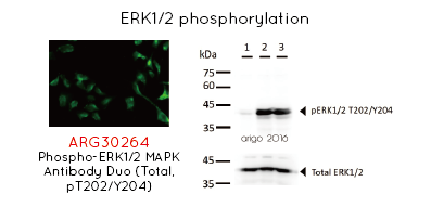
Innate immunity sense danger with toll-like receptors. Once infected, toll-like receptors will start sending downstream signals to activate NFkB signaling pathway, including phosphorylation of p65, JNK and ERK1/2. The activated proteins then transcriptionally turn on genes responsible for inflammatory responses and T cell stimulation. |
||
 |
 |
Cells undergoing apoptosis |
 |
||
|
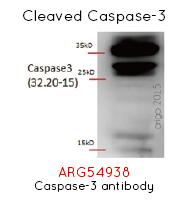
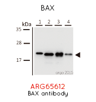
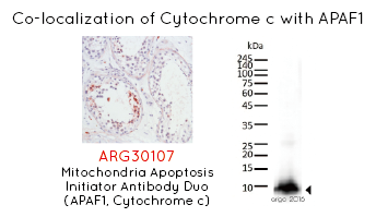
Apoptosis is a process of programmed cell death that occurs in multicellular organism. Intracellular or extracellular death signals kick-start apoptosis signaling by releasing cytochrome-c from mitochondria through BAX. The released cytochrome-c binds to APAF-1 and cleave caspase 9. Finally, caspase-3 and PARP will be cleaved and the cells officially go into apoptosis and die. |
||
 |
 |
When the cells proliferate |
|
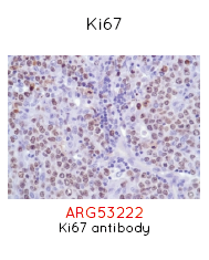
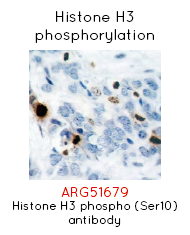
When cells are ready to proliferate, they will start expressing genes responsible for the cell cycle. Ki67 is used as a marker for cell proliferation, and it will be expressed throughout all phases (G1, S, G2, M). Phosphorylated Histone H3 at Ser-10 is restricted to cells undergoing mitosis at M phase. |
||
 |
When cell cycle |
|
|
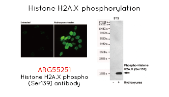
One of the main reasons for cell cycle arrest is DNA damage. Ionizing radiation, UV-light or other chemical agents causes DNA damage and result in the phosphorylation of H2A.X at Ser139 almost immediately. H2A.X is responsible for DNA repair mechanism, as well as DNA fragmentation during apoptosis. |
||
 |
 |
Cells undergoing autophagy |
|
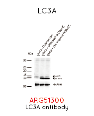
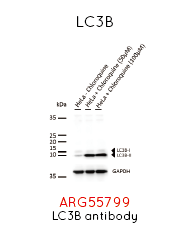
In contrast to apoptosis, autophagy is another destructive mechanism in a cell that disassemble unnecessary or dysfunctional cellular components through a regulated process. Targeted cellular constituents are enclosed by double-membraned vesicle called autophagosome. LC3A and LC3B are processed and bound to PE to form autophagosome before they are fused to lysosome and breakdown the enclosed components. |
||
 |
 |
Tumor undergoing |
|
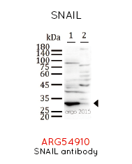
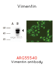
Metastasis is the spread of cancer from one organ to the other parts of body, mainly through the blood or lymphatic circulation. Before the tumor metastasize, they will express high level of SNAIL protein to down-regulate E-cadherin as they detach from the primary site. As they migrate to new site, the protein level of Vimentin, a type III intermediate filament, is also increased. It is believed that the expression of Vimentin is important for mesenchymal cell shape and motile behavior during migration. |
||
