ARG55374
anti-IDH1 antibody
anti-IDH1 antibody for Flow cytometry,ICC/IF,IHC-Formalin-fixed paraffin-embedded sections,Western blot and Human,Mouse,Rat
Cancer antibody; Metabolism antibody; Signaling Transduction antibody
Overview
| Product Description | Rabbit Polyclonal antibody recognizes IDH1 |
|---|---|
| Tested Reactivity | Hu, Ms, Rat |
| Predict Reactivity | Bov |
| Tested Application | FACS, ICC/IF, IHC-P, WB |
| Host | Rabbit |
| Clonality | Polyclonal |
| Isotype | IgG |
| Target Name | IDH1 |
| Antigen Species | Human |
| Immunogen | KLH-conjugated synthetic peptide corresponding to aa. 116-143 (Center) of Human IDH1. |
| Conjugation | Un-conjugated |
| Alternate Names | IDPC; EC 1.1.1.42; Cytosolic NADP-isocitrate dehydrogenase; IDP; HEL-S-26; HEL-216; Isocitrate dehydrogenase [NADP] cytoplasmic; IDH; PICD; IDCD; NADP; Oxalosuccinate decarboxylase |
Application Instructions
| Application Suggestion |
|
||||||||||
|---|---|---|---|---|---|---|---|---|---|---|---|
| Application Note | * The dilutions indicate recommended starting dilutions and the optimal dilutions or concentrations should be determined by the scientist. | ||||||||||
| Positive Control | HepG2 |
Properties
| Form | Liquid |
|---|---|
| Purification | This antibody is prepared by Saturated Ammonium Sulfate (SAS) precipitation followed by dialysis against PBS. |
| Buffer | PBS and 0.09% (W/V) Sodium azide |
| Preservative | 0.09% (W/V) Sodium azide |
| Storage Instruction | For continuous use, store undiluted antibody at 2-8°C for up to a week. For long-term storage, aliquot and store at -20°C or below. Storage in frost free freezers is not recommended. Avoid repeated freeze/thaw cycles. Suggest spin the vial prior to opening. The antibody solution should be gently mixed before use. |
| Note | For laboratory research only, not for drug, diagnostic or other use. |
Bioinformation
| Database Links | |
|---|---|
| Gene Symbol | IDH1 |
| Gene Full Name | isocitrate dehydrogenase 1 (NADP+), soluble |
| Background | Isocitrate dehydrogenases catalyze the oxidative decarboxylation of isocitrate to 2-oxoglutarate. These enzymes belong to two distinct subclasses, one of which utilizes NAD(+) as the electron acceptor and the other NADP(+). Five isocitrate dehydrogenases have been reported: three NAD(+)-dependent isocitrate dehydrogenases, which localize to the mitochondrial matrix, and two NADP(+)-dependent isocitrate dehydrogenases, one of which is mitochondrial and the other predominantly cytosolic. Each NADP(+)-dependent isozyme is a homodimer. The protein encoded by this gene is the NADP(+)-dependent isocitrate dehydrogenase found in the cytoplasm and peroxisomes. It contains the PTS-1 peroxisomal targeting signal sequence. The presence of this enzyme in peroxisomes suggests roles in the regeneration of NADPH for intraperoxisomal reductions, such as the conversion of 2, 4-dienoyl-CoAs to 3-enoyl-CoAs, as well as in peroxisomal reactions that consume 2-oxoglutarate, namely the alpha-hydroxylation of phytanic acid. The cytoplasmic enzyme serves a significant role in cytoplasmic NADPH production. Alternatively spliced transcript variants encoding the same protein have been found for this gene. [provided by RefSeq, Sep 2013] |
| Cellular Localization | Cytoplasm. Peroxisome |
| Highlight | Related products: Isocitrate Dehydrogenase antibodies; Isocitrate Dehydrogenase ELISA Kits; Anti-Rabbit IgG secondary antibodies; Related news: TCA intermediate fumarate promotes mitobiogenesis |
| Research Area | Cancer antibody; Metabolism antibody; Signaling Transduction antibody |
| Calculated MW | 47 kDa |
| PTM | Acetylation at Lys-374 dramatically reduces catalytic activity. |
Images (4) Click the Picture to Zoom In
-
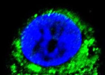
ARG55374 anti-IDH1 antibody ICC/IF image
Immunofluorescence: HepG2 cells stained with ARG55374 anti-IDH1 antibody (green). DAPI (blue) for nuclear staining.
-
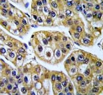
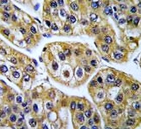
ARG55374 anti-IDH1 antibody IHC-P image
Immunohistochemistry: Formalin-fixed and paraffin-embedded Human hepatocarcinoma tissue stained with ARG55374 anti-IDH1 antibody.
-
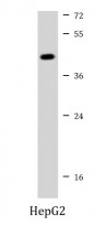
ARG55374 anti-IDH1 antibody WB image
Western blot: 35 µg of HepG2 cell lysate stained with ARG55374 anti-IDH1 antibody at 1:1000 dilution.
-
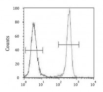
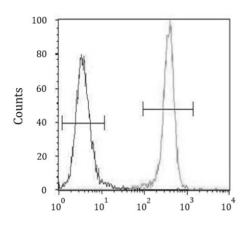
ARG55374 anti-IDH1 antibody FACS image
Flow Cytometry: 293 cells stained with ARG55374 anti-IDH1 antibody (right histogram) or without primary antibody control (left histogram), followed by incubation with FITC labelled secondary antibody.