ARG42740
anti-IDH1 antibody
anti-IDH1 antibody for Flow cytometry,ICC/IF,IHC-Frozen sections,IHC-Formalin-fixed paraffin-embedded sections,Western blot and Human,Mouse,Rat
Overview
| Product Description | Rabbit Polyclonal antibody recognizes IDH1 |
|---|---|
| Tested Reactivity | Hu, Ms, Rat |
| Predict Reactivity | Hm |
| Tested Application | FACS, ICC/IF, IHC-Fr, IHC-P, WB |
| Host | Rabbit |
| Clonality | Polyclonal |
| Isotype | IgG |
| Target Name | IDH1 |
| Antigen Species | Human |
| Immunogen | Synthetic peptide corresponding to aa. 381-413 of Human IDH1. (KGLPNVQRSDYLNTFEFMDKLGENLKIKLAQAK) |
| Conjugation | Un-conjugated |
| Alternate Names | IDPC; EC 1.1.1.42; Cytosolic NADP-isocitrate dehydrogenase; IDP; HEL-S-26; HEL-216; Isocitrate dehydrogenase [NADP] cytoplasmic; IDH; PICD; IDCD; NADP; Oxalosuccinate decarboxylase |
Application Instructions
| Application Suggestion |
|
||||||||||||
|---|---|---|---|---|---|---|---|---|---|---|---|---|---|
| Application Note | IHC-P: Antigen Retrieval: Heat mediation was performed in Citrate buffer (pH 6.0) for 20 min. * The dilutions indicate recommended starting dilutions and the optimal dilutions or concentrations should be determined by the scientist. |
||||||||||||
| Observed Size | 47 kDa |
Properties
| Form | Liquid |
|---|---|
| Purification | Affinity purification with immunogen. |
| Buffer | 0.2% Na2HPO4, 0.9% NaCl, 0.05% Sodium azide and 5% BSA. |
| Preservative | 0.05% Sodium azide |
| Stabilizer | 5% BSA |
| Concentration | 0.5 mg/ml |
| Storage Instruction | For continuous use, store undiluted antibody at 2-8°C for up to a week. For long-term storage, aliquot and store at -20°C or below. Storage in frost free freezers is not recommended. Avoid repeated freeze/thaw cycles. Suggest spin the vial prior to opening. The antibody solution should be gently mixed before use. |
| Note | For laboratory research only, not for drug, diagnostic or other use. |
Bioinformation
| Database Links | |
|---|---|
| Gene Symbol | IDH1 |
| Gene Full Name | isocitrate dehydrogenase 1 (NADP+), soluble |
| Background | Isocitrate dehydrogenases catalyze the oxidative decarboxylation of isocitrate to 2-oxoglutarate. These enzymes belong to two distinct subclasses, one of which utilizes NAD(+) as the electron acceptor and the other NADP(+). Five isocitrate dehydrogenases have been reported: three NAD(+)-dependent isocitrate dehydrogenases, which localize to the mitochondrial matrix, and two NADP(+)-dependent isocitrate dehydrogenases, one of which is mitochondrial and the other predominantly cytosolic. Each NADP(+)-dependent isozyme is a homodimer. The protein encoded by this gene is the NADP(+)-dependent isocitrate dehydrogenase found in the cytoplasm and peroxisomes. It contains the PTS-1 peroxisomal targeting signal sequence. The presence of this enzyme in peroxisomes suggests roles in the regeneration of NADPH for intraperoxisomal reductions, such as the conversion of 2, 4-dienoyl-CoAs to 3-enoyl-CoAs, as well as in peroxisomal reactions that consume 2-oxoglutarate, namely the alpha-hydroxylation of phytanic acid. The cytoplasmic enzyme serves a significant role in cytoplasmic NADPH production. Alternatively spliced transcript variants encoding the same protein have been found for this gene. [provided by RefSeq, Sep 2013] |
| Cellular Localization | Cytoplasm. Peroxisome. [UniProt] |
| Highlight | Related products: Isocitrate Dehydrogenase antibodies; Isocitrate Dehydrogenase ELISA Kits; Anti-Rabbit IgG secondary antibodies; Related news: TCA intermediate fumarate promotes mitobiogenesis |
| Calculated MW | 47 kDa |
| PTM | Acetylation at Lys-374 dramatically reduces catalytic activity. [UniProt] |
Images (7) Click the Picture to Zoom In
-

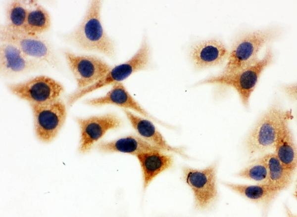
ARG42740 anti-IDH1 antibody ICC image
Immunocytochemistry: A549 cells were blocked with 10% goat serum and then stained with ARG42740 anti-IDH1 antibody at 1 µg/ml dilution, overnight at 4°C.
-
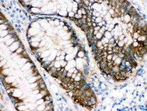
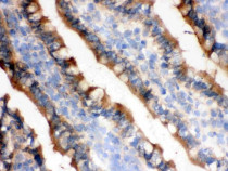
ARG42740 anti-IDH1 antibody IHC-P image
Immunohistochemistry: Paraffin-embedded Human intestinal cancer tissue. Antigen Retrieval: Heat mediation was performed in Citrate buffer (pH 6.0) for 20 min. The tissue section was blocked with 10% goat serum. The tissue section was then stained with ARG42740 anti-IDH1 antibody at 1 µg/ml dilution, overnight at 4°C.
-
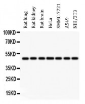
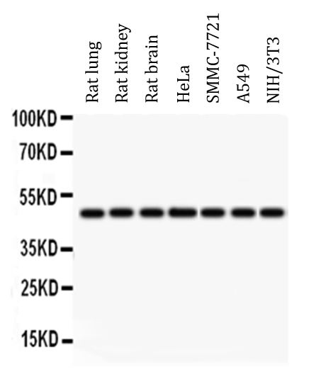
ARG42740 anti-IDH1 antibody WB image
Western blot: 50 µg of sample under reducing conditions. Rat lung, Rat kidney, Rat brain, HeLa, SMMC-7721, A549 and NIH/3T3 whole cell lysates stained with ARG42740 anti-IDH1 antibody at 0.5 µg/ml dilution, overnight at 4°C.
-
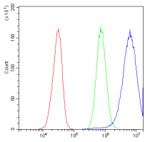
ARG42740 anti-IDH1 antibody FACS image
Flow Cytometry: HepG2 cells were blocked with 10% normal goat serum and then stained with ARG42740 anti-IDH1 antibody (blue) at 1 µg/10^6 cells for 30 min at 20°C, followed by incubation with DyLight®488 labelled secondary antibody. Isotype control antibody (green) was Rabbit IgG (1 µg/10^6 cells) used under the same conditions. Unlabelled sample (red) was also used as a control.
-
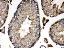
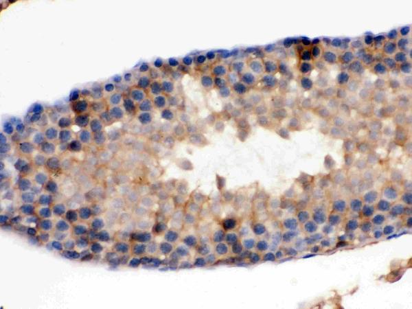
ARG42740 anti-IDH1 antibody IHC-P image
Immunohistochemistry: Paraffin-embedded Mouse testis tissue. Antigen Retrieval: Heat mediation was performed in Citrate buffer (pH 6.0) for 20 min. The tissue section was blocked with 10% goat serum. The tissue section was then stained with ARG42740 anti-IDH1 antibody at 1 µg/ml dilution, overnight at 4°C.
-

ARG42740 anti-IDH1 antibody IHC-P image
Immunohistochemistry: Paraffin-embedded Rat testis tissue. Antigen Retrieval: Heat mediation was performed in Citrate buffer (pH 6.0) for 20 min. The tissue section was blocked with 10% goat serum. The tissue section was then stained with ARG42740 anti-IDH1 antibody at 1 µg/ml dilution, overnight at 4°C.
-
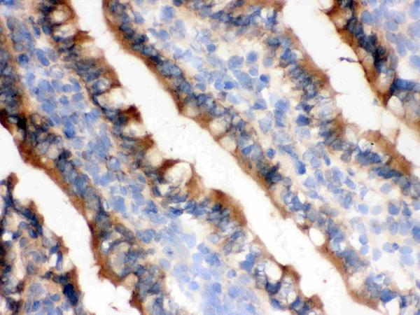
ARG42740 anti-IDH1 antibody IHC-Fr image
Immunohistochemistry: Frozen section of Rat small intestine tissue. The tissue section was blocked with 10% goat serum. The tissue section was then stained with ARG42740 anti-IDH1 antibody at 1 µg/ml dilution, overnight at 4°C.